
Page 383 - Carrahers_Polymer_Chemistry,_Eighth_Edition
P. 383
346 Carraher’s Polymer Chemistry
Cell wall Virus
A
dsRNA
Dicer
B
C
Normal single-stranded RNA
D
F
E
Cytoplasm
Single-stranded viral RNA
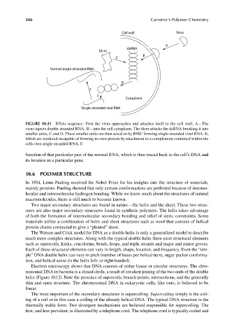
FIGURE 10.11 RNAi sequence. First the virus approaches and attaches itself to the cell wall, A—The
virus injects double-stranded RNA, B—into the cell cytoplasm. The dicer attacks the dsRNA breaking it into
smaller units, C and D. These smaller units are then acted on by RISC forming single-stranded viral RNA, E,
which are rendered incapable of forming its own protein by attachment to a complement contained within the
cells own single-stranded RNA, F.
function of that particular part of the normal RNA, which is then traced back to the cell’s DNA and
its location in a particular gene.
10.6 POLYMER STRUCTURE
In 1954, Linus Pauling received the Nobel Prize for his insights into the structure of materials,
mainly proteins. Pauling showed that only certain conformations are preferred because of intermo-
lecular and intermolecular hydrogen bonding. While we know much about the structures of natural
macromolecules, there is still much to become known.
Two major secondary structures are found in nature—the helix and the sheet. These two struc-
tures are also major secondary structures found in synthetic polymers. The helix takes advantage
of both the formation of intermolecular secondary bonding and relief of steric constraints. Some
materials utilize a combination of helix and sheet structures such as wool that consists of helical
protein chains connected to give a “pleated” skeet.
The Watson and Crick model for DNA as a double helix is only a generalized model to describe
much more complex structures. Along with the typical double helix there exist structural elements
such as supercoils, kinks, cruciforms, bends, loops, and triple strands and major and minor groves.
Each of these structural elements can vary in length, shape, location, and frequency. Even the “sim-
ple” DNA double helix can vary in pitch (number of bases per helical turn), sugar pucker conforma-
tion, and helical sense (is the helix left- or right-handed).
Electron microscopy shows that DNA consists of either linear or circular structures. The chro-
mosomal DNA in bacteria is a closed circle, a result of covalent joining of the two ends of the double
helix (Figure 10.12). Note the presence of supercoils, branch points, intersections, and the generally
thin and open structure. The chromosomal DNA in eukaryotic cells, like ours, is believed to be
linear.
The most important of the secondary structures is supercoiling. Supercoiling simply is the coil-
ing of a coil or in this case a coiling of the already helical DNA. The typical DNA structure is the
thermally stable form. Two divergent mechanisms are believed responsible for supercoiling. The
first, and less prevalent, is illustrated by a telephone cord. The telephone cord is typically coiled and
9/14/2010 3:41:19 PM
K10478.indb 346 9/14/2010 3:41:19 PM
K10478.indb 346