
Page 77 - Color Atlas of Biochemistry
P. 77
68 Biomolecules
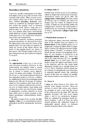
Secondary structures B. Collagen helix
Another type of helix occurs in the collagens,
In proteins, specific combinations of the dihe-
dral angles φ and ψ (see p. 66) are much more which are important constituents of the con-
nectivetissue matrix (see pp. 70, 344). The
common than others. When several succes-
sive residues adopt one of these conforma- collagen helix is left-handed,and with a pitch
of 0.96 nm and 3.3 residues per turn, it is
tions, defined secondary structures arise,
which are stabilized by hydrogen bonds ei- steeper than the α-helix. In contrast to the
α-helix, H bonds are not possible within the
ther within the peptide chain or between
neighboring chains. When a large part of a collagen helix. However, the conformation is
protein takes on a defined secondary struc- stabilized by the association of three helices
ture, the protein often forms mechanically to form a righthanded collagen triple helix
(see p. 70).
stable filaments or fibers. Structural proteins
of this type (see p. 70) usually have character-
istic amino acid compositions. C. Pleated-sheet structures
The most important secondary structural
elements of proteins are discussed here first. Two additional, almost stretched, conforma-
The illustrations only show the course of the tions of the peptide chain are known as E
peptide chain; the side chains are omitted. To pleated sheets, as the peptide planes are ar-
make thecourseof the chains clearer, the ranged like a regularly folded sheet of paper.
levels of the peptide bonds are shown as Again, H bonds can only form between neigh-
boring chains (“strands”) in pleated sheets.
blue planes. The dihedral angles of the struc-
tures shown here are also marked in diagram When the two strands run in opposite direc-
D1 on p. 67. tions (1), the structure is referred to as an
antiparallel pleated sheet (β a ). When they
runinthe same direction(2), it is a parallel
A. D-Helix pleated sheet (β p ). In both cases, the α-C
atoms occupy the highest and lowest points
The right-handed α-helix (α R )is one of the
most common secondary structures. In this in the structure, and the side chains point
conformation, the peptide chain is wound alternately straight up or straight down (see
like a screw. Each turn of the screw (the screw p. 71 C). The β a structure, with its almost lin-
ear H bonds, is energetically more favorable.
axis in shown in orange) covers approxi-
mately 3.6 amino acid residues. The pitch of In extended pleated sheets, the individual
the screw (i. e., the smallest distance between strands of the sheet are usually not parallel,
but twisted relative to one another (see p. 74).
two equivalent points) is 0.54 nm. α-Helices
are stabilized by almost linear hydrogen bonds
between the NH and CO groups of residues, D. E Turns
which are four positions apart from each an-
other in the sequence (indicated by red dots; E Turns are often found at sites where the
peptide chain changes direction. These are
see p. 6). In longer helices, most amino acid
residuesthusenter into two Hbonds.Apolar sections in which four amino acid residues
or amphipathic α-helices with five to seven are arranged in such a way that the course
turns often serve to anchor proteins in bio- of the chain reverses by about 180° into the
logical membranes (transmembrane helices; opposite direction. The two turns shown
(types I and II) are particularly frequent.
see p. 214).
The mirror image of the α R helix, the left- Both are stabilized by hydrogen bonds be-
handed D-helix (α L ), is rarely found in nature, tween residues 1 and 4. β Turns are often
although it would be energetically “permissi- located between the individual strands of
ble.” antiparallel pleated sheets, or between
strands of pleated sheets and α helices.
Koolman, Color Atlas of Biochemistry, 2nd edition © 2005 Thieme
All rights reserved. Usage subject to terms and conditions of license.