
Page 106 - Computational Modeling in Biomedical Engineering and Medical Physics
P. 106
Electrical activity of the heart 95
Electrophysiology insights
The heart has the role of an electric generator and circuit for the transmission of cyclical
voltage pulses, as local cell transmembrane voltage alternates its state between resting and
active or polarized and depolarized conditions. The muscular tissue of the atria and ventri-
cles, the conductive tissue (Hiss branches and the Purkinje network) and the specialized
pacemaker cells [SA and atrioventricular (AV) nodes] are its main parts. Electrical depolari-
zation waves initiated at the SA node diffuse through the conductive cellular network up
to the muscular cells in a remarkably orderly manner and drive the cyclical contrac-
tion relaxation of the myocardium, ensuring the blood circulation through the body.
In the cyclic functioning of a healthy heart two successive sequences are distinguish-
able: (1) the systole, or active phase, which consists in the atria contraction followed by
the ventricles contraction, while the blood is forced through the atrioventricular route
and circulatory pathways, and (2) the diastole, or passive phase, during which the myo-
cardium relaxes and the four heart chambers are filled with blood. This happens under
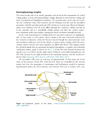
the electrical signals that are generated and spread throughout a complex and redundant
conduction system, made as shown by Fig. 4.1 of the SA node [located between the
superior vena cava (SVC) and the right atrium (RA)], the internodal atrial pathways, the
AV node (located in the right posterior of the interatrial septum), and the electrocon-
ductive tissue (Ganong, 2005; Keener and Sneyd, 2009; Katz, 2011).
SA pacemaker cells cover an oval area of approximately 15-mm long and 5-mm
wide, at the junction of the SVC with the RA. They are controlled by the nervous
system but have the properties of automatism and rhythmicity needed to generate
depolarization pulses, by self-excitation, and maintain their tact to initiate each cycle
Figure 4.1 Conduction system of the heart. ,https://commons.wikimedia.org/w/index.php?
curid 5 29922595..