
Page 262 - Computational Modeling in Biomedical Engineering and Medical Physics
P. 262
Hyperthermia and ablation 251
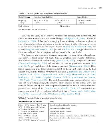
Table 8.1 Electromagnetic field and thermal therapy methods.
Medical therapy Hyperthermia and ablation Laser ablation
2
2
2
Frequency 10 kHz 10 MHz 10 MHz 10 GHz 10 THz 10 PHz
range (RF) (MW) (light)
EMF radiation Nonionizing
EMF, Electromagnetic field; RF, radio frequency.
The Joule heat injury on the tumor is determined by the local, total electric work, the
tumor microenvironment, and the tumor biology (Nikfarjam et al., 2005a), as cited in
Habash et al., (2006). Although the underlying thermosensitivity mechanisms imply com-
plex cellular and subcellular interactions within tumor tissue, it is the cell membrane seems
to be the most vulnerable to heat injury. In vitro (Dickson and Calderwood, 1980)and
in vivo (Overgaard and Overgaard, 1972)[as citedin Habash et al. (2006)] studies evidence
that tumor cells are killed at temperatures lower than for the normal cells.
The hyperthermia application triggers a progression in tissue damage through sev-
eral factors: induced cancer cell death through apoptosis, microvascular destruction,
and ischemia reperfusion related injury (Brown et al., 1992), Kupffer cells activation
(Tsutsui and Nishiguchi, 2014) and alteration of cytokine peptides expression (Welc
et al., 2012), and modulation of the immune response (Baronzio et al., 2006). These
factors depend on tissue temperatures produced by the applied total power, the heat
exhaust rate, and the thermal properties of the tissue (Christensen and Durney, 1981;
Dewhirst et al., 2003a; Haemmerich and Laeseke, 2005; Haemmerich et al., 2005;
Nikfarjam et al., 2005b; Osepchuk, Petersen, 2001; Seegenschmiedt and Vernon,
1995; Vander Vorst et al., 2006). The underlying relationships between thermal expo-
sure and the pending damage, the collateral effects endured by the normal tissue, and
methods for converting one time-temperature protocol to time at standardized tem-
perature are reviewed in Dewhirst et al. (2003b). Table 8.2 summarizes the
temperature-related effects produced to biological tissues (Germer et al., 1998; Habash
et al., 2006; Haemmerich et al., 2005; Lepock, 2005; Stauffer, 2005).
Table 8.2 Thermal effects, temperature range, duration.
Temperature range and duration Effects
Below 250 C for more than 10 min Complete cellular killing by freezing
30 C 39 C for no specific duration Hyperthermic cellular death
40 C 46 C for 30 60 min
47 C 50 C for more than 10 min Protein denaturation, necrosis
Over 50 C after B2 min Necrosis, cell death
60 C 140 C for seconds Protein denaturation, membrane rupture, cell stricture, ablation
100 C 300 C for seconds Vaporization of extracellular steam vacuoles
Over 300 C for fraction of a second Carbonization