Page 264 - Computational Modeling in Biomedical Engineering and Medical Physics
P. 264
Hyperthermia and ablation  253


                   temperature reaches the critical value (T h respectively T c ), and (2) the temperature
                   exceeds (hyperthermia) or decreases below (cryoablation) the critical value for a certain
                   time (t h , respectively t c ).
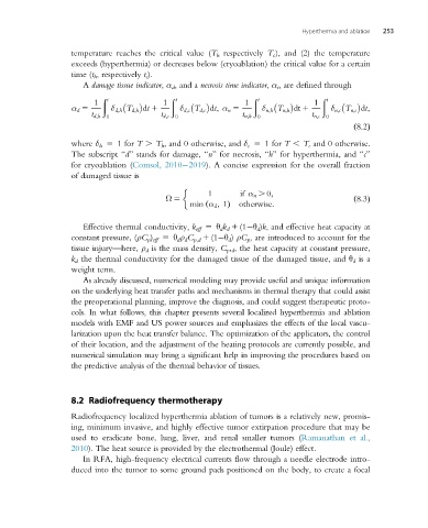
                      A damage tissue indicator, α d , and a necrosis time indicator, α t , are defined through

                           ð t              ð t                  ð t              ð t
                         1                1                    1                1
                   α d 5     δ d;h T d;h dt 1  δ d;c T d;c dt; α n 5  δ n;h T n;h dt 1  δ n;c T n;c dt;
                        t d;h 0           t d;c  0            t n;h 0           t n;c  0
                                                                                          ð8:2Þ
                   where δ h 5 1 for T . T h , and 0 otherwise, and δ c 5 1 for T , T c and 0 otherwise.
                   The subscript “d” stands for damage, “n” for necrosis, “h” for hyperthermia, and “c”
                   for cryoablation (Comsol, 2010 2019). A concise expression for the overall fraction
                   of damaged tissue is


                                                     1       if α n . 0;
                                           Ω 5                                            ð8:3Þ
                                                 min α d ; 1ð  Þ otherwise:
                      Effective thermal conductivity, k eff 5 θ d k d 1 (1 θ d )k, and effective heat capacity at
                   constant pressure, (ρC p ) eff 5 θ d ρ d C p,d 1 (1 θ d ) ρC p , are introduced to account for the
                   tissue injury—here, ρ d is the mass density, C p , d , the heat capacity at constant pressure,
                   k d the thermal conductivity for the damaged tissue of the damaged tissue, and θ d is a
                   weight term.
                      As already discussed, numerical modeling may provide useful and unique information
                   on the underlying heat transfer paths and mechanisms in thermal therapy that could assist
                   the preoperational planning, improve the diagnosis, and could suggest therapeutic proto-
                   cols. In what follows, this chapter presents several localized hyperthermia and ablation
                   models with EMF and US power sources and emphasizes the effects of the local vascu-
                   larization upon the heat transfer balance. The optimization of the applicators, the control
                   of their location, and the adjustment of the heating protocols are currently possible, and
                   numerical simulation may bring a significant help in improving the procedures based on
                   the predictive analysis of the thermal behavior of tissues.


                   8.2 Radiofrequency thermotherapy

                   Radiofrequency localized hyperthermia ablation of tumors is a relatively new, promis-
                   ing, minimum invasive, and highly effective tumor extirpation procedure that may be
                   used to eradicate bone, lung, liver, and renal smaller tumors (Ramanathan et al.,
                   2010). The heat source is provided by the electrothermal (Joule) effect.
                      In RFA, high-frequency electrical currents flow through a needle electrode intro-
                   duced into the tumor to some ground pads positioned on the body, to create a focal

