
Page 268 - Computational Modeling in Biomedical Engineering and Medical Physics
P. 268
Hyperthermia and ablation 257
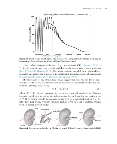
Figure 8.2 Blood vessels classification. After Feijóo, 2000. Computational methods in biology. In:
Proceedings of the 2nd Summer School LNCC/MCT, Petrópolis, Brazil.
Using online imaging techniques (e.g., unenhanced CT) (Zagoria, 2004), a
“LeVeen” array of electrodes is positioned close to the tumor using a trocar guideway
(Fig. 8.1B and C)(Boston, 2013). The tumor volume, modeled by an ellipsoid here,
is located in a region that is prone to its proliferation through genesis and angiogenesis
(Maeshima and Makino, 2010; Osteaux and Jeanmart, 1979).
The time scales of the physics that concur suggest that from the first law perspec-
tive, the RF-EMF heat and electric work interactions are consistently modeled as elec-
trokinetic (Morega et al., 2020)
rU 2σrVÞ 5 0; ð8:4Þ
ð
where V is the electric potential and σ is the electrical conductivity. Dirichlet
boundary conditions are set for the kidney surface (ground) and for the electrode tips
(V 5 22 V), which provide the required power level for a successful ablation (Fig. 8.3,
left). The inlet arterial velocity (uniform profile) is 0.1 m/s and a uniform pressure
profile is set for the vein outlet.
Figure 8.3 Boundary conditions for the RF ablation model. RF, Radio frequency (Morega et al., 2020).