
Page 4 - Academic Press Encyclopedia of Physical Science and Technology 3rd Molecular Biology
P. 4
P1: GQQ Revised Pages
Encyclopedia of Physical Science and Technology EN002G-90 May 17, 2001 20:42
542 Cell Death (Apoptosis)
APOPTOSIS, or programed cell death, plays an impor- and Kerr in 1972. In the necrotic process, swelling of cells
tant role in the development of organisms and in the main- precedes their explosion and results in the release of in-
tenance of homeostasis. The failure of apoptotic programs tracellular components that may be toxic to other cells.
causes various diseases. So far, many genes regulating In apoptosis, the dying cells exhibit nuclear and cytoplas-
apoptosis have been identified and the molecular mecha- mic condensation, fragmentation of cell bodies, chromo-
nism of apoptosis is being clarified. In this article, I will somal DNA fragmentation into nucleosomal units, loss
discuss cell death elicited through death receptors. of mitochondrial function, and alterations of cell mem-
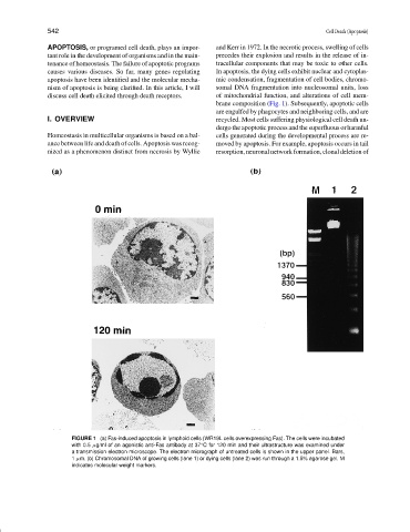
brane composition (Fig. 1). Subsequently, apoptotic cells
are engulfed by phagocytes and neighboring cells, and are
I. OVERVIEW recycled. Most cells suffering physiological cell death un-
dergo the apoptotic process and the superfluous or harmful
Homeostasis in multicellular organisms is based on a bal- cells generated during the developmental process are re-
ance between life and death of cells. Apoptosis was recog- moved by apoptosis. For example, apoptosis occurs in tail
nized as a phenomenon distinct from necrosis by Wyllie resorption, neuronal network formation, clonal deletion of
FIGURE 1 (a) Fas-induced apoptosis in lymphoid cells (WR19L cells overexpressing Fas). The cells were incubated
with 0.5 µg/ml of an agonistic anti-Fas antibody at 37 C for 120 min and their ultrastructure was examined under
◦
a transmission electron microscope. The electron micrograph of untreated cells is shown in the upper panel. Bars,
1 µm. (b) Chromosomal DNA of growing cells (lane 1) or dying cells (lane 2) was run through a 1.5% agarose gel. M
indicates molecular weight markers.