
Page 229 - Environmental Nanotechnology Applications and Impacts of Nanomaterials
P. 229
214 Principles and Methods
as well as their state of aggregation, dispersability, and solubility. These
properties under aqueous conditions play important roles in particle
interactions with cells, binding to the cell membrane, cellular uptake, and
subcellular distribution. Asufficient number of particles need to be taken
up to induce adverse biological effects. Material composition, reactive sur-
face groups/chemicals, and crystallinity are key determinants in the
ability of nanoparticles to generate ROS and oxidant injury [42].
Cell-free assays to determine ROS production
There are numerous assays that can be performed to test the inherent
properties of NM to produce ROS under abiotic conditions (Table 6.3).
Electron spin resonance (ESR) detects unpaired electrons in any given
sample. Ascorbate and spin-trapping agents such as 5,5-dimethyl-1-
pyrroline-N-oxide (DMPO) can be used to detect free oxygen radicals
[43]. ESR assessment is usually performed at room temperature
with a quartz flat cell and a spectrometer that records the ESR spectra.
The spectra are characterized by the shape of the absorption curve, the
position of the resonance field, the line width, and the area under the
absorption curve. Software programs, such as EPRWare, can help to
superimpose and quantify the ratio of superoxide and hydroxyl radicals
on their DMPO spin adducts, thereby allowing one to calculate the free
radical concentration [43].
The dithiothreitol (DTT) assay can be used to measure the presence
of redox cycling organic chemicals such as quinones on the surface of
ambient ultrafine particles [44]. Quinones are capable of capturing
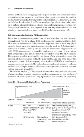
TABLE 6.3 Hierarchical Oxidative Stress Responses
High GSH/GSSG ratio Low GSH/GSSG ratio
Level of
oxidative
stress
Methods Normal Tier 1 Tier 2 Tier 3
Cell defense Pro-inflammation Mitochondria effects, apoptosis
in vitro HO-1 WB Cytokine ELISA Mitochondrial ∆Ψm (DiOC )
6
2+
2+
Phase II enzymes Phospho-JNK WB [Ca ] : Fluo-3; [Ca ] : Rhod-2
i
m
(Real-time PCR) Apoptosis: Annexin V/PI
Nrf-2 WB Caspase 3 activation
DNA fragmentation(BrdU-FITC)
in vivo HO-1 Luc Mice BAL cytokines Cell damage (LDH)
Inflammatory cells Immunohistochemistry
Histology (active caspase-3)
Adapted from [31].