
Page 231 - Environmental Nanotechnology Applications and Impacts of Nanomaterials
P. 231
216 Principles and Methods
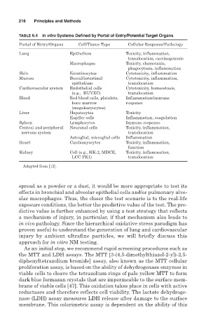
TABLE 6.4 In vitro Systems Defined by Portal of Entry/Potential Target Organs
Portal of Entry/Organs Cell/Tissue Type Cellular Response/Pathology
Lung Epithelium Toxicity, inflammation,
translocation, carcinogenesis
Macrophages Toxicity, chemotaxis,
phagocytosis, inflammation
Skin Keratinocytes Cytotoxicity, inflammation
Mucosa Buccal/intestinal Cytotoxicity, inflammation,
epithelium translocation
Cardiovascular system Endothelial cells Cytotoxicity, homeostasis,
(e.g., HUVEC) translocation
Blood Red blood cells, platelets, Inflammation/immune
bone marrow response
(megakaryocytes)
Liver Hepatocytes Toxicity
Kupffer cells Inflammation, coagulation
Spleen Lymphocytes Immune response
Central and peripheral Neuronal cells Toxicity, inflammation,
nervous system translocation
Astroglial, microglial cells Inflammation
Heart Cardiomyocytes Toxicity, inflammation,
function
Kidney Cell (e.g., HK-2, MDCK, Toxicity, inflammation,
LCC-PK1) translocation
Adapted from [12].
spread as a powder or a dust, it would be more appropriate to test its
effects in bronchial and alveolar epithelial cells and/or pulmonary alve-
olar macrophages. Thus, the closer the test scenario is to the real-life
exposure conditions, the better the predictive value of the test. The pre-
dictive value is further enhanced by using a test strategy that reflects
a mechanism of injury, in particular, if that mechanism also leads to
in vivo pathology. Since the hierarchical oxidative stress paradigm has
proven useful to understand the generation of lung and cardiovascular
injury by ambient ultrafine particles, we will briefly discuss this
approach for in vitro NM testing.
As an initial step, we recommend rapid screening procedures such as
the MTT and LDH assays. The MTT [3-(4,5-dimethylthiazol-2-yl)-2,5-
diphenyltetrazolium bromide] assay, also known as the MTT cellular
proliferation assay, is based on the ability of dehydrogenase enzymes in
viable cells to cleave the tetrazolium rings of pale yellow MTT to form
dark blue formazan crystals that are impermeable to the surface mem-
brane of viable cells [47]. This oxidation takes place in cells with active
reductases and therefore reflects cell viability. The lactate dehydroge-
nase (LDH) assay measures LDH release after damage to the surface
membrane. This colorimetric assay is dependent on the ability of this