
Page 236 - Environmental Nanotechnology Applications and Impacts of Nanomaterials
P. 236
Principles and Procedures to Assess Nanomaterial Toxicity 221
A transgenic line was established that provides a rapid and noninvasive
way to characterize in vivo HO-1 expression in response to systemic agents.
Agents that elicit oxidative stress (e.g., CdCl 2 , heme, and other metallo-
porphyrins) have been used in these mice for in vivo assays [53, 54]. Avari-
ety of HO-1 expression profiles have been demonstrated based on the
stimulus, method of administration, and time since administration [54].
For instance, intravenous administration of CdCl 2 results in a luciferase
signal originating primarily from the liver, while peritoneal administra-
tion of heme yields a response in the liver, spleen, and other abdominal
sites [55]. Both CdCl 2 and heme induce this response based on their abil-
ities to generate ROS [55]. We have successfully tested the ROS generat-
ing ability of ambient UFP using this animal model and believe it could
be helpful for launching nanotoxicology studies. Similar imaging models
exist to study the activation of the NF- B signaling cascade.
Nanosensors: Sensitive Probes
for the Biodetection of ROS
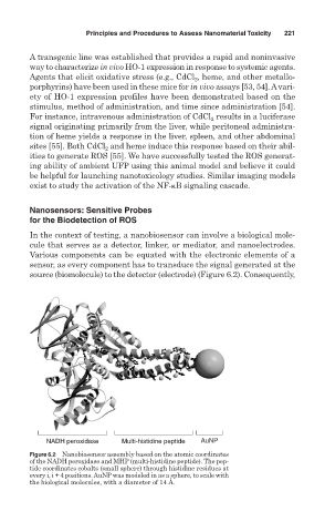
In the context of testing, a nanobiosensor can involve a biological mole-
cule that serves as a detector, linker, or mediator, and nanoelectrodes.
Various components can be equated with the electronic elements of a
sensor, as every component has to transduce the signal generated at the
source (biomolecule) to the detector (electrode) (Figure 6.2). Consequently,
NADH peroxidase Multi-histidine peptide AuNP
Figure 6.2 Nanobiosensor assembly based on the atomic coordinates
of the NADH peroxidase and MHP (multi-histidine peptide). The pep-
tide coordinates cobalts (small sphere) through histidine residues at
every i, i + 4 positions. AuNP was modeled in as a sphere, to scale with
the biological molecules, with a diameter of 14 Å.