
Page 452 - Environmental Nanotechnology Applications and Impacts of Nanomaterials
P. 452
Toxicological Impacts of Nanomaterials 429
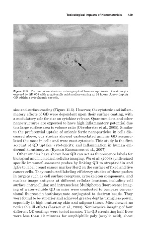
Figure 11.5 Transmission electron micrograph of human epidermal keratinocyte
exposed to QD 655 with a carboxylic acid surface coating at 24 hours. Arrow depicts
QD within a cytoplasmic vacuole.
size and surface coating (Figure 11.5). However, the cytotoxic and inflam-
matory effects of QD were dependent upon their surface coating, with
a modulatory role for size on cytokine release. Quantum dots and other
nanostructures are expected to have high inflammatory potential due
to a large surface area to volume ratio (Oberdorster et al., 2005). Similar
to the preferential uptake of anionic ferric nanoparticles in cells dis-
cussed above, our studies showed carboxylated anionic QD accumu-
lated the most in cells and were most cytotoxic. This study is the first
account of QD uptake, cytotoxicity, and inflammation in human epi-
dermal keratinocytes (Ryman-Rasmussen et al., 2007).
Other studies have shown how QD can act as fluorescence labels for
biological and biomedical cellular imaging. Wu et al. (2003) synthesized
specific immunofluorescent probes by linking QD to streptavidin and
IgGs to label breast cancer marker Her2 on the surface of fixed and live
cancer cells. They conducted labeling efficiency studies of these probes
in targets such as cell surface receptors, cytoskeleton components, and
nuclear image antigens at different cellular locations, including cell
surface, intracellular, and intranuclear. Multiphoton fluorescence imag-
ing of water-soluble QD in mice were conducted to compare conven-
tional fluorescein isothiocyanate conjugated to dextran beads. They
were found to be superior and achieved greater depths using less power,
especially in high scattering skin and adipose tissue. Mice showed no
noticeable ill effects (Larson et al., 2003). Noninvasive imaging of four
different QD coatings were tested in mice. The QD circulating half-lives
were less than 12 minutes for amphiphilic poly (acrylic acid), short