
Page 455 - Environmental Nanotechnology Applications and Impacts of Nanomaterials
P. 455
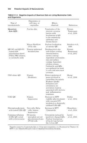
432   Potential Impacts of Nanomaterials

        TABLE 11.3  Negative Impacts of Quantum Dots on Living Mammalian Cells
        and Organisms
                           Organisms, or
           Type of          cell types, or      Effects
        nanomaterials        organelles         observed        References
        Quantum           Porcine skin    Penetration of the  Ryman-
         dots (QD)                         stratum corneum     Rasmussen
                                           barrier with        et al., 2006
                                           localization of QD
                                           in the underlying
                                           epidermal and
                                           dermal layers
                          Mouse fibroblast  Nuclear localization  Bruchez et al.,
                           (3T3) cells     of cationic QD      1998
        QD 565 and QD 655   Human epidermal  Nondependent size   Ryman-
         coated with       keratinocytes   and surface coating  Rasmussen
         polyethylene glycol               internalization;    et al., 2007
         (PEG), PEG-amines,                surface coating–
         or carboxylic acids               dependent cytotoxicity;
                                           size and surface
                                           coating–dependent
                                           cytokine release.
                                           Carboxylic acid QDs
                                           accumulated the most
                                           in cells and were most
                                           cytotoxic.
        PEG-silane-QD     Human           Down-regulation of  Zhang
                           fibroblasts     genes involved in   et al., 2006
                                           controlling the mitotic
                                           M-phase spindle
                                           formation and
                                           cytokinesis; no
                                           activation of genes
                                           associated with immune
                                           and  inflammatory
                                           responses
        CdSe QD           Human           Decrease in MTT     Derfus
                           hepatocytes     viability after treatment  et al., 2004
                                           with uncoated UV
                                           irradiated or chemically
                                           oxidized CdSe QD
        Mercaptoundecanoic   Vero cells, HeLa  Increase in MTT  Shiosahara
         acid-coated CdSe QD  cells, human  Cytotoxicity       et al., 2004
                           hepatocytes
        Cadmium tellerium   N9 murine     Cytotoxicity induced by  Lovri ´ c
         (CdTe) QD coated   microglial and rat  positively charged QDs.  et al., 2005
         with cysteineamine  pheochromocytoma  Nuclear localization
                           (PC12) cells    of the 2.3 nm but not
                                           the 5.2 nm QD
          Negative impacts of nanomaterials on living microorganisms are illustrated in Chapter 12.
          MTT: 3-[4,5-dimethylthiazol-2-yl]-2,5-diphenyltetrazolium bromide.