
Page 55 - Fiber Bragg Gratings
P. 55
36 Chapter 2 Photosensitivity and Photosensitization of Optical Fibers
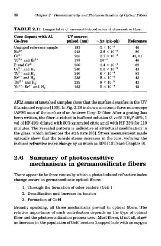
TABLE 2.1: League table of rare-earth-doped silica photosensitive fiber.
Core dopant with Al, UV source:
Ge-free pulsed (nm) —An (pk-pk) Reference
Undoped reference sample 193 5 x 10 46
Eu 2+ 248 2.5 x 1(T 5 80
3+ 4
Ce 265 3.7 x 10~ 43, 81
3+ 3+ 4
Yb and Er 193 10~ 46
P and Ce 3+ 266 1.4 X 10~ 4 82
3+ 3
Ce and H 2 240 1.5 X 10~ 43
3+ 4
Tb and H 2 240 6 X 10~ 83
3+ 5
Er and H 2 235 5 X HT 43
3+ 5
Tm and H 2 235 8 X 10~ 43
3+ 3+ 4
Yb : Er and H 2 193 5 x 10~ 43
AFM scans of unetched samples show that the surface densifies in the UV
illuminated regions [100]. In Fig. 2.13 is shown an atomic force microscope
(AFM) scan of the surface of an Andrew Corp. D-fiber. After a grating has
been written, the fiber is etched in buffered solution (3 vol% NH 4F 40%, 1
vol of HF 49% diluted with 50% saturated citric acid) with HF 25% for 110
minutes. The revealed pattern is indicative of structural modification in
the glass, which influences the etch rate [88]. Stress measurement made
optically show that the tensile stress increases in the core, reducing the
induced refractive index change by as much as 30% [101] (see Chapter 9).
2.6 Summary of photosensitive
mechanisms in germanosilicate fibers
There appear to be three routes by which a photo-induced refractive index
change occurs in germanosilicate optical fibers:
1. Through the formation of color centers (GeE')
2. Densification and increase in tension
3. Formation of GeH
Broadly speaking, all three mechanisms prevail in optical fibers. The
relative importance of each contribution depends on the type of optical
fiber and the photosensitization process used. Most fibers, if not all, show
an increase in the population of GeE' centers (trapped hole with an oxygen