
Page 142 - Fundamentals of Light Microscopy and Electronic Imaging
P. 142
DOUBLE REFRACTION IN CRYSTALS 125
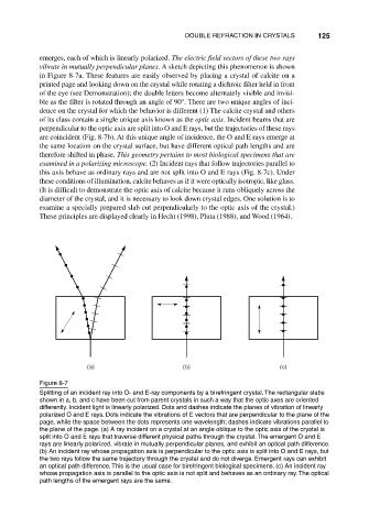
emerges, each of which is linearly polarized. The electric field vectors of these two rays
vibrate in mutually perpendicular planes. A sketch depicting this phenomenon is shown
in Figure 8-7a. These features are easily observed by placing a crystal of calcite on a
printed page and looking down on the crystal while rotating a dichroic filter held in front
of the eye (see Demonstration); the double letters become alternately visible and invisi-
ble as the filter is rotated through an angle of 90°. There are two unique angles of inci-
dence on the crystal for which the behavior is different (1) The calcite crystal and others
of its class contain a single unique axis known as the optic axis. Incident beams that are
perpendicular to the optic axis are split into O and E rays, but the trajectories of these rays
are coincident (Fig. 8-7b). At this unique angle of incidence, the O and E rays emerge at
the same location on the crystal surface, but have different optical path lengths and are
therefore shifted in phase. This geometry pertains to most biological specimens that are
examined in a polarizing microscope. (2) Incident rays that follow trajectories parallel to
this axis behave as ordinary rays and are not split into O and E rays (Fig. 8-7c). Under
these conditions of illumination, calcite behaves as if it were optically isotropic, like glass.
(It is difficult to demonstrate the optic axis of calcite because it runs obliquely across the
diameter of the crystal, and it is necessary to look down crystal edges. One solution is to
examine a specially prepared slab cut perpendicularly to the optic axis of the crystal.)
These principles are displayed clearly in Hecht (1998), Pluta (1988), and Wood (1964).
(a) (b) (c)
Figure 8-7
Splitting of an incident ray into O- and E-ray components by a birefringent crystal. The rectangular slabs
shown in a, b, and c have been cut from parent crystals in such a way that the optic axes are oriented
differently. Incident light is linearly polarized. Dots and dashes indicate the planes of vibration of linearly
polarized O and E rays. Dots indicate the vibrations of E vectors that are perpendicular to the plane of the
page, while the space between the dots represents one wavelength; dashes indicate vibrations parallel to
the plane of the page. (a) A ray incident on a crystal at an angle oblique to the optic axis of the crystal is
split into O and E rays that traverse different physical paths through the crystal. The emergent O and E
rays are linearly polarized, vibrate in mutually perpendicular planes, and exhibit an optical path difference.
(b) An incident ray whose propagation axis is perpendicular to the optic axis is split into O and E rays, but
the two rays follow the same trajectory through the crystal and do not diverge. Emergent rays can exhibit
an optical path difference. This is the usual case for birefringent biological specimens. (c) An incident ray
whose propagation axis is parallel to the optic axis is not split and behaves as an ordinary ray. The optical
path lengths of the emergent rays are the same.