
Page 58 - Fundamentals of Light Microscopy and Electronic Imaging
P. 58
EFFECTS OF LIGHT ON LIVING CELLS 41
/4 high-index
layer
Dielectric reflector
/4 low stack
index layer Cavity spacer layer
/2 thick
Dielectric reflector
stack
Coupling layer
Dielectric reflector
stack
Cavity spacer layer
/2 thick
Dielectric reflector
stack
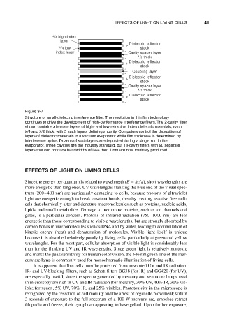
Figure 3-7
Structure of an all-dielectric interference filter. The revolution in thin film technology
continues to drive the development of high-performance interference filters. The 2-cavity filter
shown contains alternate layers of high- and low-refractive index dielectric materials, each
/4 and /2 thick, with 5 such layers defining a cavity. Computers control the deposition of
layers of dielectric materials in a vacuum evaporator while film thickness is determined by
interference optics. Dozens of such layers are deposited during a single run in the
evaporator. Three cavities are the industry standard, but 18-cavity filters with 90 separate
layers that can produce bandwidths of less than 1 nm are now routinely produced.
EFFECTS OF LIGHT ON LIVING CELLS
Since the energy per quantum is related to wavelength (E hc/ ), short wavelengths are
more energetic than long ones. UV wavelengths flanking the blue end of the visual spec-
trum (200–400 nm) are particularly damaging to cells, because photons of ultraviolet
light are energetic enough to break covalent bonds, thereby creating reactive free radi-
cals that chemically alter and denature macromolecules such as proteins, nucleic acids,
lipids, and small metabolites. Damage to membrane proteins, such as ion channels and
gates, is a particular concern. Photons of infrared radiation (750–1000 nm) are less
energetic than those corresponding to visible wavelengths, but are strongly absorbed by
carbon bonds in macromolecules such as DNA and by water, leading to accumulation of
kinetic energy (heat) and denaturation of molecules. Visible light itself is unique
because it is absorbed relatively poorly by living cells, particularly at green and yellow
wavelengths. For the most part, cellular absorption of visible light is considerably less
than for the flanking UV and IR wavelengths. Since green light is relatively nontoxic
and marks the peak sensitivity for human color vision, the 546 nm green line of the mer-
cury arc lamp is commonly used for monochromatic illumination of living cells.
It is apparent that live cells must be protected from unwanted UV and IR radiation.
IR- and UV-blocking filters, such as Schott filters BG38 (for IR) and GG420 (for UV),
are especially useful, since the spectra generated by mercury and xenon arc lamps used
in microscopy are rich in UV and IR radiation (for mercury, 30% UV, 40% IR, 30% vis-
ible; for xenon, 5% UV, 70% IR, and 25% visible). Phototoxicity in the microscope is
recognized by the cessation of cell motility and the arrest of organelle movement; within
3 seconds of exposure to the full spectrum of a 100 W mercury arc, amoebae retract
filopodia and freeze, their cytoplasm appearing to have gelled. Upon further exposure,