
Page 66 - Fundamentals of Light Microscopy and Electronic Imaging
P. 66
OBJECT-IMAGE MATH 49
• a f: No real image exists that can be projected on a screen. If the eye is placed
behind the lens, a virtual image is perceived on the far side of the lens.
• a f: The image distance b is infinite, so no image exists that can be projected on
a screen. We used this condition previously to determine the focal length of a lens,
only in reverse: Parallel beams of light from an “infinitely distant” object converge
at the focal length of the lens. This is the case for image formation in a telescope.
For the condition that a f, a real image is always formed. The unique domains for
this condition are as follows:
•2f a f: A real magnified image is formed. This arrangement is used for pro-
ducing the first real image in a microscope.
• a 2f: This is a specialized case. Under this condition, b 2f also. A real image is
formed, but there is no magnification and M 1.
• a 2f: A real demagnified image is formed and M 1.
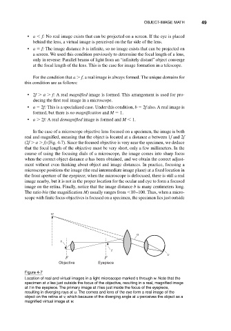
In the case of a microscope objective lens focused on a specimen, the image is both
real and magnified, meaning that the object is located at a distance a between 1f and 2f
(2f a f) (Fig. 4-7). Since the focused objective is very near the specimen, we deduce
that the focal length of the objective must be very short, only a few millimeters. In the
course of using the focusing dials of a microscope, the image comes into sharp focus
when the correct object distance a has been obtained, and we obtain the correct adjust-
ment without even thinking about object and image distances. In practice, focusing a
microscope positions the image (the real intermediate image plane) at a fixed location in
the front aperture of the eyepiece; when the microscope is defocused, there is still a real
image nearby, but it is not in the proper location for the ocular and eye to form a focused
image on the retina. Finally, notice that the image distance b is many centimeters long.
The ratio b/a (the magnification M) usually ranges from 10–100. Thus, when a micro-
scope with finite focus objectives is focused on a specimen, the specimen lies just outside
w
u
s
t
v
F F
Objective Eyepiece
Figure 4-7
Location of real and virtual images in a light microscope marked s through w. Note that the
specimen at s lies just outside the focus of the objective, resulting in a real, magnified image
at t in the eyepiece. The primary image at t lies just inside the focus of the eyepiece,
resulting in diverging rays at u. The cornea and lens of the eye form a real image of the
object on the retina at v, which because of the diverging angle at u perceives the object as a
magnified virtual image at w.