
Page 92 - Handbook of Biomechatronics
P. 92
Sensors and Transducers 87
of attention. The alpha wave indicates the person is awake with his or her
eyes closed. The beta wave signifies mental activity and attention. The theta
and delta waves are correlated with drowsiness, sleep, or a pathological con-
dition (Fernandez et al., 2014).
Although easy to setup, since the EEG electrodes are placed externally,
and relatively inexpensive, EEG is often criticized for its low resolution.
EEG is useful in recording brain signals, but it has been difficult interpret
the individual electrode signals.
To determine if you have built Jacob a better system, you decide to have
Jacob do two EEG recordings, one where he pedals a stationary bike while
using his passive leg, and another where he pedals using his new leg. After
the test, you compare the EEG recordings, focusing specifically on beta
waves to see which device required greater mental focus.
6.2 Electrocardiogram
Since the heart is a muscle, as the four chambers contact and relax, the mus-
cle responses release voltage signals, similar to standard muscle contraction.
The heart also has special Purkinje fibers, or “pacemaker” cells, which
initiate electrical signals regulate systematic chamber contraction. An elec-
trocardiogram, EKG or ECG, is the transducer system which is used to
measure the voltage output of the heart (Purves et al., 2008b).
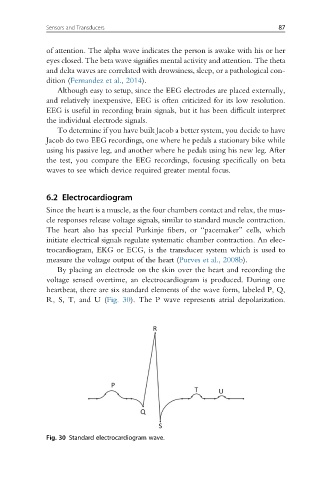
By placing an electrode on the skin over the heart and recording the
voltage sensed overtime, an electrocardiogram is produced. During one
heartbeat, there are six standard elements of the wave form, labeled P, Q,
R, S, T, and U (Fig. 30). The P wave represents atrial depolarization.
Fig. 30 Standard electrocardiogram wave.