
Page 88 - Handbook of Biomechatronics
P. 88
Sensors and Transducers 83
For Jacob’s prosthetic system, you decide again to change how he varies
the knee resistance. You decide to use two surface EMG preamplifiers
mounted in the socket for the residual limb of his leg. You configure the
system such that when he flexes his residual limb muscles, the resistance of
the knee goes down. When he extends his residual limb muscles, the knee
resistance goes up.
5.3 Intramuscular EMG
Instead of collecting noisy EMG signals from the surface, EMG signals can be
collected by placing EMG electrodes directly into the muscle. There are two
methods of collecting this intramuscular EMG. The first is to insert an intra-
muscular needle electrode into the muscle site of interest. Depending on the
type of needle electrode used, a ground needle electrode is often required.
By collecting the signals directly from the muscles, the signal tends to be
cleaner. Although useful for short-term experiments, overtime the body
tends to reject the wire electrodes. Also, in areas such as the forearm, where
multiple muscles are closely located, specific muscle targeting can be difficult.
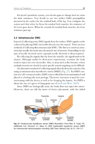
An alternative method of collecting signals directly from the muscles is by
using an intramuscular myoelectric sensor (IMES) (Fig. 27). An IMES con-
sists of a self-contained surface EMG sensor which has been miniaturized and
placed in a biologically inert package. Electronic resonance is used for com-
municating with the device as well as for charging the battery. An IMES is
about the size of a piece of long-grain rice (Farrell and Weir, 2007).
Since IMES are biologically inert, the body does not reject the sensor.
However, there are still the issues of device placement, with the added
Fig. 27 Intramuscular myoelectric sensor (IMES) illustration. From Weir, R., Troyk, P.R.,
DeMichele, G.A., Schorsch, J.F., Maas, H., 2009. Implantable myoelectric sensors (IMES)
for intramuscular electromyogram recording. IEEE Trans. Biomed. Eng. 56 (1), 159–171.