
Page 85 - Handbook of Biomechatronics
P. 85
80 Jeff Christenson
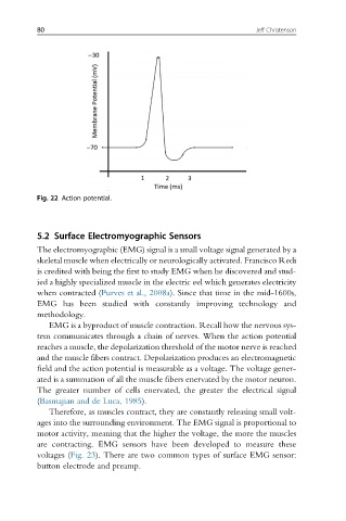
Fig. 22 Action potential.
5.2 Surface Electromyographic Sensors
The electromyographic (EMG) signal is a small voltage signal generated by a
skeletal muscle when electrically or neurologically activated. Francisco Redi
is credited with being the first to study EMG when he discovered and stud-
ied a highly specialized muscle in the electric eel which generates electricity
when contracted (Purves et al., 2008a). Since that time in the mid-1600s,
EMG has been studied with constantly improving technology and
methodology.
EMG is a byproduct of muscle contraction. Recall how the nervous sys-
tem communicates through a chain of nerves. When the action potential
reaches a muscle, the depolarization threshold of the motor nerve is reached
and the muscle fibers contract. Depolarization produces an electromagnetic
field and the action potential is measurable as a voltage. The voltage gener-
ated is a summation of all the muscle fibers enervated by the motor neuron.
The greater number of cells enervated, the greater the electrical signal
(Basmajian and de Luca, 1985).
Therefore, as muscles contract, they are constantly releasing small volt-
ages into the surrounding environment. The EMG signal is proportional to
motor activity, meaning that the higher the voltage, the more the muscles
are contracting. EMG sensors have been developed to measure these
voltages (Fig. 23). There are two common types of surface EMG sensor:
button electrode and preamp.