
Page 84 - Handbook of Biomechatronics
P. 84
Sensors and Transducers 79
5.1 Neuromuscular Anatomy
The neuromuscular system consists of three main elements: the central ner-
vous system, nerves, and muscles. The central nervous system is comprised
of the brain and spinal cord. Muscles include not only standard muscles such
as the biceps and calves, but also muscles in the heart, lungs, eyes, etc.
Efferent biological signals, signals which are generated in the central nervous
system and travel to periphery systems, originate in the brain or spinal cord
and travel through thousands of nerves until it reaches its final destination
and causes the body to respond. Along the way, that signal passes through
the spinal cord, down nerve cords, to nerve centers and into the target organ
or muscle. Afferent biological signals, those generated in the periphery
systems and sent to the central nervous system, travel a similar but opposite
path to the brain (Bolton, 2003e).
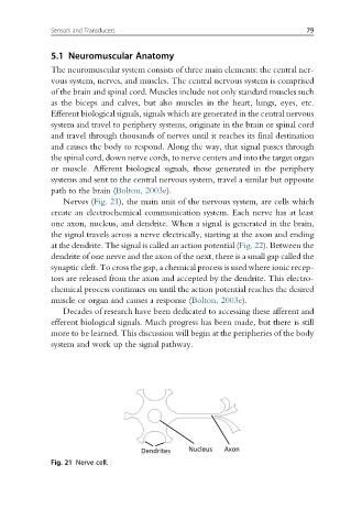
Nerves (Fig. 21), the main unit of the nervous system, are cells which
create an electrochemical communication system. Each nerve has at least
one axon, nucleus, and dendrite. When a signal is generated in the brain,
the signal travels across a nerve electrically, starting at the axon and ending
at the dendrite. The signal is called an action potential (Fig. 22). Between the
dendrite of one nerve and the axon of the next, there is a small gap called the
synaptic cleft. To cross the gap, a chemical process is used where ionic recep-
tors are released from the axon and accepted by the dendrite. This electro-
chemical process continues on until the action potential reaches the desired
muscle or organ and causes a response (Bolton, 2003e).
Decades of research have been dedicated to accessing these afferent and
efferent biological signals. Much progress has been made, but there is still
more to be learned. This discussion will begin at the peripheries of the body
system and work up the signal pathway.
Fig. 21 Nerve cell.