
Page 18 - Handbook of Electronic Assistive Technology
P. 18
Chapter 1 • Basic Neurosciences With Relevance to Electronic Assistive Technology 5
massive growth of the brain after we are born that differentiates us from other mammals.
Unlike wildebeest that need for obvious reasons to speed off across the plains straight after
birth, the complex reorganisation that occurs in the human brain postnatally increases the
potential complexity of sensory, motor and in particular cognitive interaction and reason-
ing that we have. As such we are unusual because we have the largest brain of all animals,
in comparison to body weight, and most of this growth occurs postnatally.
But this time of rapid brain growth is also a period of great risk for the development of a
number of neurological problems. Each structure in the nervous system has a period when
it is particularly sensitive to the normal influences of the chemical, physical and physi-
ological environment surrounding the foetus in the developing womb, such as intrinsic
blood supply and external oxygenation, nutrients, growth factors and hormones. If these
are compromised at critical points, then focal or global development of the brain can be
compromised. It’s a process fraught with the possibilities of grey matter structures devel-
oping wrongly or white matter pathways going haywire.
So far so easy? If you look in clinic at the anatomical picture provided by a magnetic res-
onance image (MRI) of the brain, it is relatively identical in a 2-year-old, a 12-year-old and
a 32-year-old. However, with the advent of functional neuroimaging (in particular fMRI)
we have been able to look at how the fine wiring of the system develops rather than just
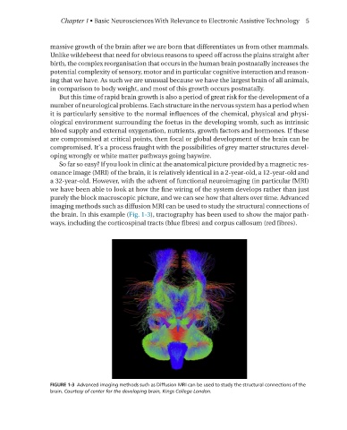
purely the block macroscopic picture, and we can see how that alters over time. Advanced
imaging methods such as diffusion MRI can be used to study the structural connections of
the brain. In this example (Fig. 1-3), tractography has been used to show the major path-
ways, including the corticospinal tracts (blue fibres) and corpus callosum (red fibres).
FIGURE 1-3 Advanced imaging methods such as Diffusion MRI can be used to study the structural connections of the
brain� Courtesy of center for the developing brain, Kings College London.