
Page 211 - Handbook of Properties of Textile and Technical Fibres
P. 211
188 Handbook of Properties of Textile and Technical Fibres
domains (Rising et al., 2005, 2006; Motriuk-Smith et al., 2005) and are covalently
linked via cysteine bridges in their termini (Sponner et al., 2005b; Humenik et al.,
2011; Hayashi and Lewis, 2000).
In the repetitive region, a glycine (G)-rich repeat followed by an alanine (A) block
appears consecutively about 100 times, making up 60% of the entire sequence
(Hinman and Lewis, 1992). Alanine-rich domains have been assigned specific second-
ary structures such as b-sheet, 3 1 -helix, and b-spiral that form crystalline structures in
the fiber (Xu and Lewis, 1990; Simmons et al., 1994, 1996; K€ ummerlen et al., 1996;
Hayashi et al., 1999), while glycine-rich regions are believed to be more amorphous
(Simmons et al., 1996; Hayashi et al., 1999). Therefore the proportion of alanine
blocks that determine the level of crystallinity in the fiber is correlated to the strength
and stiffness of the fiber, and glycine-rich regions contribute to the extensibility and
flexibility of the fiber. However, it is noteworthy that there are considerable numbers
of studies arguing that the structure of the poly-Gly regions may be semiordered in a
3 1 -helix (K€ ummerlen et al., 1996) or type 1 b-turn (van Beek et al., 2000; Jelinski
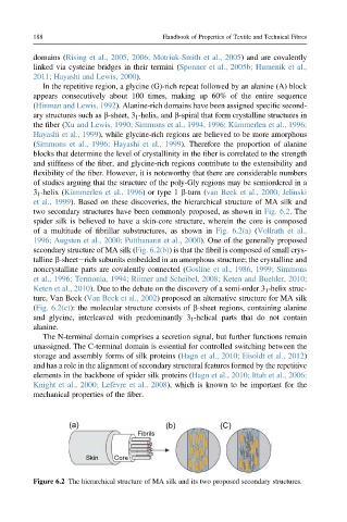
et al., 1999). Based on these discoveries, the hierarchical structure of MA silk and
two secondary structures have been commonly proposed, as shown in Fig. 6.2. The
spider silk is believed to have a skin-core structure, wherein the core is composed
of a multitude of fibrillar substructures, as shown in Fig. 6.2(a) (Vollrath et al.,
1996; Augsten et al., 2000; Putthanarat et al., 2000). One of the generally proposed
secondary structure of MA silk (Fig. 6.2(b)) is that the fibril is composed of small crys-
talline b-sheeterich subunits embedded in an amorphous structure; the crystalline and
noncrystalline parts are covalently connected (Gosline et al., 1986, 1999; Simmons
et al., 1996; Termonia, 1994; R€ omer and Scheibel, 2008; Keten and Buehler, 2010;
Keten et al., 2010). Due to the debate on the discovery of a semi-order 3 1 -helix struc-
ture, Van Beek (Van Beek et al., 2002) proposed an alternative structure for MA silk
(Fig. 6.2(c)): the molecular structure consists of b-sheet regions, containing alanine
and glycine, interleaved with predominantly 3 1 -helical parts that do not contain
alanine.
The N-terminal domain comprises a secretion signal, but further functions remain
unassigned. The C-terminal domain is essential for controlled switching between the
storage and assembly forms of silk proteins (Hagn et al., 2010; Eisoldt et al., 2012)
and has a role in the alignment of secondary structural features formed by the repetitive
elements in the backbone of spider silk proteins (Hagn et al., 2010; Ittah et al., 2006;
Knight et al., 2000; Lef evre et al., 2008), which is known to be important for the
mechanical properties of the fiber.
(a) (b) (C)
Fibrils
Skin Core
Figure 6.2 The hierarchical structure of MA silk and its two proposed secondary structures.