
Page 113 - Handbook of Surface Improvement and Modification
P. 113
108 Surface Tension and Wetting
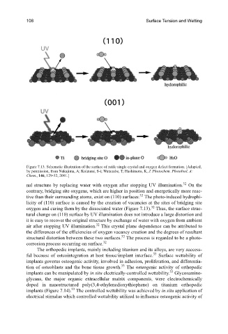
Figure 7.13. Schematic illustration of the surface of rutile single crystal and oxygen defect formation. [Adapted,
by permission, from Nakajima, A; Koizumi, S-i; Watanabe, T; Hashimoto, K, J. Photochem. Photobiol. A:
Chem., 146, 129-32, 2001.]
32
nal structure by replacing water with oxygen after stopping UV illumination. On the
contrary, bridging site oxygens, which are higher in position and energetically more reac-
32
tive than their surrounding atoms, exist on (110) surfaces. The photo-induced hydrophi-
licity of (110) surface is caused by the creation of vacancies at the sites of bridging site
32
oxygen and curing them by the dissociated water (Figure 7.13). Thus, the surface struc-
tural change on (110) surface by UV illumination does not introduce a large distortion and
it is easy to recover the original structure by exchange of water with oxygen from ambient
32
air after stopping UV illumination. This crystal plane dependence can be attributed to
the differences of the efficiencies of oxygen vacancy creation and the degrees of resultant
32
structural distortion between these two surfaces. The process is regarded to be a photo-
32
corrosion process occurring on surface.
The orthopedic implants, mainly including titanium and its alloys, are very success-
33
ful because of osteointegration at host tissue/implant interface. Surface wettability of
implants governs osteogenic activity, involved in adhesion, proliferation, and differentia-
33
tion of osteoblasts and the bone tissue growth. The osteogenic activity of orthopedic
33
implants can be manipulated by in situ electrically-controlled wettability. Glycosamino-
glycans, the major organic extracellular matrix components, were electrochemically
doped in nanostructured poly(3,4-ethylenedioxythiophene) on titanium orthopedic
33
implants (Figure 7.14). The controlled wettability was achieved by in situ application of
electrical stimulus which controlled wettability utilized to influence osteogenic activity of