
Page 97 - Human Inspired Dexterity in Robotic Manipulation
P. 97
Approaching Human Hand Dexterity Through Highly Biomimetic Design 95
side of the finger bones. On the other side, after passing through the carpal
tunnel, the flexor tendons travel through a series of pulley-like tendon
sheaths grown onto the palmar side of the bones and eventually insert at
the base of the DIP and PIP joints. The collaborative motions of the two
tendon groups make fluent hand movement possible.
The large muscle groups that directly connect to the central branch of the
flexor and extensor tendons are called extrinsic muscles. Most of them orig-
inate from the elbow and have muscle bellies located in the forearm. Differ-
ent groups of muscles help to realize a subset of hand movements ranging
from twisting the wrist to bending the fingers. However, there also exist
several smaller muscle groups called intrinsic muscles. The majority of these
small muscles start from the wrist of the hand and connect to the thinner
branches of the extensor tendons of each finger near the MCP joint. Most
of their muscle bellies are slim enough to reside in the gap between the two
adjacent metacarpal bones. One important function of these intrinsic
muscles is to stabilize the finger joints during various hand activities.
Most of our daily tasks involving hand motions require the contraction of
strong muscles connecting to the flexor tendons. However, during this pro-
cess, the extensor tendons also work as a breaking system that constantly reg-
ulates the motion of fingers. The functionality of the breaking system relies
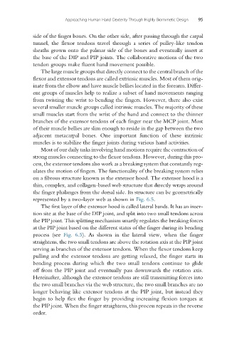
on a fibrous structure known as the extensor hood. The extensor hood is a
thin, complex, and collagen-based web structure that directly wraps around
the finger phalanges from the dorsal side. Its structure can be geometrically
represented by a two-layer web as shown in Fig. 6.5.
The first layer of the extensor hood is called lateral bands. It has an inser-
tion site at the base of the DIP joint, and split into two small tendons across
the PIP joint. This splitting mechanism smartly regulates the breaking forces
at the PIP joint based on the different status of the finger during its bending
process (see Fig. 6.5). As shown in the lateral view, when the finger
straightens, the two small tendons are above the rotation axis at the PIP joint
serving as branches of the extensor tendons. When the flexor tendons keep
pulling and the extensor tendons are getting relaxed, the finger starts its
bending process during which the two small tendons continue to glide
off from the PIP joint and eventually pass downwards the rotation axis.
Hereinafter, although the extensor tendons are still transmitting forces into
the two small branches via the web structure, the two small branches are no
longer behaving like extensor tendons at the PIP joint, but instead they
begin to help flex the finger by providing increasing flexion torques at
the PIP joint. When the finger straightens, this process repeats in the reverse
order.