
Page 237 - Industrial Ventilation Design Guidebook
P. 237
1 98 CHAPTER 5 PHYSIOLOGICAL AND TOXICOLOGICAL CONSIDERATIONS
vapor between the airway wall and the inspiratory or expiratory air-
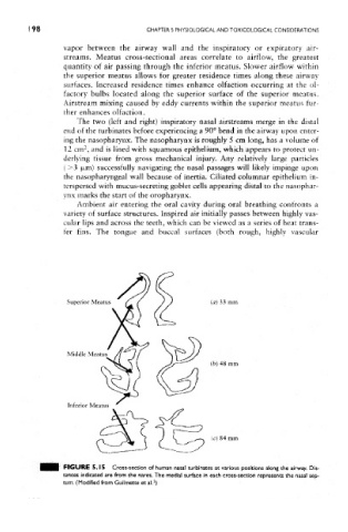
streams, Meatus cross-sectional areas correlate to airflow, the greatest
quantity of air passing through the inferior meatus. Slower airflow within
the superior meatus allows for greater residence times along these airway
surfaces. Increased residence times enhance olfaction occurring at the ol-
factory bulbs located along the superior surface of the superior meatus.
Airstream mixing caused by eddy currents within the superior meatus fur-
ther enhances olfaction.
The two (left and right) inspiratory nasal airstreams merge in the distal
end of the turbinates before experiencing a 90° bend in the airway upon enter-
ing the nasopharynx. The nasopharynx is roughly 5 cm long, has a volume of
2
12 cm , and is lined with squamous epithelium, which appears to protect un-
derlying tissue from gross mechanical injury. Any relatively large particles
{ >3 |xm) successfully navigating the nasal passages will likely impinge upon
the nasopharyngeal wall because of inertia. Ciliated columnar epithelium in-
terspersed with mucus-secreting goblet cells appearing distal to the nasophar-
ynx marks the start of the oropharynx.
Ambient air entering the oral cavity during oral breathing confronts a
variety of surface structures. Inspired air initially passes between highly vas-
cular lips and across the teeth, which can be viewed as a series of heat trans-
fer fins. The tongue and buccal surfaces (both rough, highly vascular
FIGURE 5.15 Cross-section of human nasal turbinates at various positions along the airway. Dis-
tances indicated are from the nares. The medial surface in each cross-section represents the nasal sep-
2
tum. (Modified from Guilmette et al. )