
Page 241 - Industrial Ventilation Design Guidebook
P. 241
202 CHAPTER 5 PHYSIOLOGICAL AND TOXiCOLOGlCAL CONSIDERATIONS
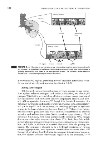
FIGURE 5.16 Depiction of representative airway cross-section at various points (trachea, bronchi,
and pulmonary airway) along the respiratory tract showing common cell types. Note how mucus gel is
generally presumed to form sheets in the more proximal airways. The pulmonary airway depiction
includes both a section of respiratory bronchi and an alveolus.
more vulnerable) regions, permitting more of these fine participates to set-
tle in distal airways by sedimentation (see Section 5.2.7).
Airway Surface Liquid
ASL lining the airway luminal surface serves to protect airway epithe-
lium against airborne pathogens and toxins, desiccation, and abrupt pH
changes. This fluid is secreted along all airway surfaces except portions of
the extrathoracic and respiratory airways (respiratory bronchi and alve-
9 10
oli). ASL composition is unclear, ' though it is theorized to consist of a
periciliary layer composed mainly of water and various ions approximately
10
5-7 jxm in depth and the epiphase, an overlying gel layer of hydrated
11 13
mucins in the form of droplets, sheets, or blankets " (Fig, 5.16). Epithe-
lial cells control periciliary fluid water and ion concentration by chloride
secretion and sodium absorption. Solids constitute approximately 5% of
periciliary fluid mass, with water comprising the remaining 95%, though
disease can raise solids concentration above 10%. Periciliary fluid solids
include glycoproteins, proteins, peptides, glycosaminoglycans, immunoglo-
bins, and lipids in addition to materials deposited from the passing air-
stream. The epiphase is thought to be a hydrogel consisting of various
14 15
complex glycoproteins, with hydration controlled by a Donnan effect, '
Control of periciliary fluid hydration is a complex interaction of evapora-
10 16 11 17 18
tion, ' osmotic pressure differentials regulated by ion transport, - -