Page 244 - Industrial Ventilation Design Guidebook
P. 244
5.2 HUMAN RESPIRATORY TRACT PHYSIOLOGY                                    205

                      Intravascular blood pressure drops as a function of arterial and arteriole
                  diameter to such an extent that capillary walls can consist of a single layer of
                  endothelial cells. Capillaries, with diameters of 6-8 (xm, transport blood close
                  enough (roughly 20-30 jxm) to cells throughout the body to allow gas (O 2 and
                  CO 2), heat, nutrient and waste, and water exchange between blood and cells
                  via diffusion. Interstitial tissue containing collagen fibers, basement mem-
                  branes, elastin, and water supports capillary endothelial cells and provides ad-
                  ditional tensile strength. Capillaries merge to form venules, which in turn
                  merge to form veins. These low-pressure components of the cardiovascular
                  system—capillaries, venules, and veins—transport deoxygenated blood from
                  the capillaries to the right atrium of the heart via the largest vein, the inferior
                  vena cava.
                       Blood supplying conducting airway tissues derives from large bron-
                  chial arteries branching off either the aorta or intercostal arteries. These
                  vessels also supply blood to the visceral pleura, regional nerves and lymph
                  nodes, and vascular walls of the pulmonary arteries and veins. Bronchial
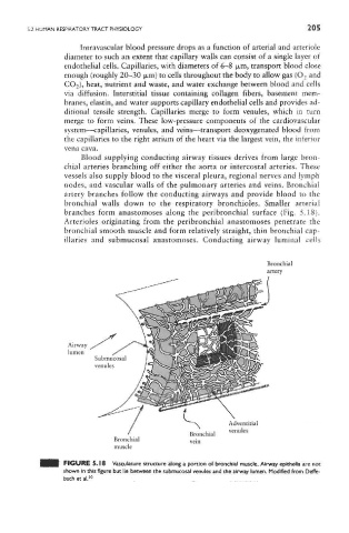
                  artery branches follow the conducting airways and provide blood to the
                  bronchial walls down to the respiratory bronchioles. Smaller arterial
                  branches form anastomoses along the peribronchial surface (Fig. 5.18).
                  Arterioles originating from the peribronchial anastomoses penetrate the
                  bronchial smooth muscle and form relatively straight, thin bronchial cap-
                  illaries and submucosal anastomoses. Conducting airway luminal cells


                  FIGURE 5.18 Vasculature structure along a portion of bronchial muscle. Airway epithelia are not
                  shown in this figure but lie between the submucosal venules and the airway lumen. Modified from Deffe-
                  bach et al 20

