Page 348 - Introduction to Paleobiology and The Fossil Record
P. 348

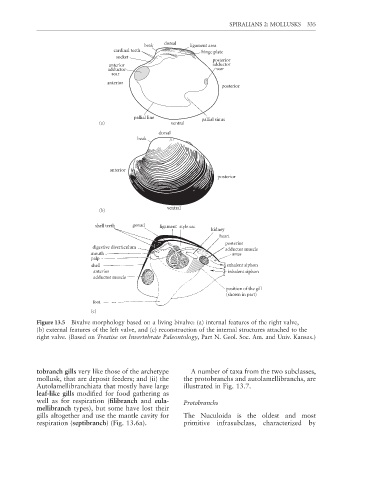
SPIRALIANS 2: MOLLUSKS  335


                                                       dorsal
                                                beak           ligament area
                                      cardinal teeth               hinge plate
                                       socket
                                                                      posterior
                                     anterior                         adductor
                                    adductor                            scar
                                     scar
                                    anterior
                                                                         posterior


                                             pallial line          pallial sinus
                                 (a)                     ventral
                                                     dorsal
                                              beak


                                     anterior
                                                                        posterior


                                                        ventral
                                 (b)

                                shell teeth  gonad   ligament style sac
                                                                      kidney
                                                                         heart
                                                                           posterior
                               digestive diverticulum
                                                                           adductor muscle
                               mouth                                         anus
                               palp
                               shell                                       exhalent siphon
                                anterior                                    inhalent siphon
                                adductor muscle
                                                                           position of the gill
                                                                           (shown in part)
                               foot
                               (c)
             Figure 13.5  Bivalve morphology based on a living bivalve: (a) internal features of the right valve,
             (b) external features of the left valve, and (c) reconstruction of the internal structures attached to the
             right valve. (Based on Treatise on Invertebrate Paleontology, Part N. Geol. Soc. Am. and Univ. Kansas.)


             tobranch gills very like those of the archetype   A number of taxa from the two subclasses,
             mollusk, that are deposit feeders; and (ii) the   the protobranchs and autolamellibranchs, are
             Autolamellibranchiata that mostly have large    illustrated in Fig. 13.7.

             leaf-like gills modified for food gathering as
             well as for respiration (fi libranch and  eula-  Protobranchs
             mellibranch types), but some have lost their
             gills altogether and use the mantle cavity for   The Nuculoida is the oldest and most
             respiration (septibranch) (Fig. 13.6a).         primitive infrasubclass, characterized by
   343   344   345   346   347   348   349   350   351   352   353