
Page 156 - Materials Chemistry, Second Edition
P. 156
143
2.4. The Amorphous State
Figure 2.98. Crystal structure of hydroxyapatite, projected on the (x,y) plane. Reproduced with
permission from Nature, 1964, 204, 1050.
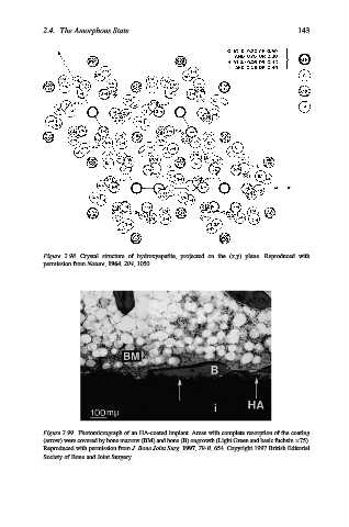
Figure 2.99. Photomicrograph of an HA-coated implant. Areas with complete resorption of the coating
(arrow) were covered by bone marrow (BM) and bone (B) ongrowth (Light Green and basic fuchsin 75).
Reproduced with permission from J. Bone Joint Surg. 1997, 79-B, 654. Copyright 1997 British Editorial
Society of Bone and Joint Surgery.