
Page 40 - Nanotechnology an introduction
P. 40
composite structure essentially consisting of mineral nanoplates cemented together by a protein matrix [87]. The exact mechanism of their
formation has yet to be elucidated but presumably is essentially an extracellular process. Bone is apparently constructed on similar principles,
although it has a more sophisticated hierarchical structure.
4.1.3. Cells
This section considers two themes: (1) a nano-object small enough to penetrate inside a cell; and (2) the response of a supported cell to
nanostructured features of its substratum (support).
Penetration of Nano-Objects into the Cell's Interior
It has been a long-standing frustration of electrophysiologists that the electrodes typically used to probe electrical activity in the interior of the brain
are gigantic in comparison with the size of neurons and dendrites, hence observations not only imply gross damage due to initial installation of the
electrode, but also that signals are recorded simultaneously from dozens of neurons. The availability of carbon nanotubes, which can be excellent
electrical conductors (Section 9.2), offers the possibility of creating an electrode considerably smaller than a single cell. This has the potential to
revolutionize experimental neurophysiology. The main practical obstacle is the difficulty of manipulating the nanotubes: they must be attached to
some kind of back plate. If the nano-object is freely floating, such as the drug delivery nanoparticle already referred to, the main problem is
controlling uptake. The tiniest nanoparticles might be able to enter the cell through some of the larger transmembrane channels. Larger
nanoparticles may be engulfed (e.g., by macrophages) as if they were viruses. They may also become wrapped by the outer lipid membranes of
the cell, as has been observed in a model system [117]. The penetration of a drug-bearing nanoparticle into the cell's interior may be precisely the
goal of the particle designer, but nanoparticles used in other applications for cosmetic purposes, for example as ultraviolet-absorbing sunscreen
applied to the skin, may, by similar mechanisms, also end up inside the cell where they may have toxic consequences. The investigation of these
consequences constitutes what is currently the most active part of the field of nanotoxicology (Section 4.3).
The Response of a Cell to External Nanostructured Features
The fundamental premise engendering interest in living cells interacting with nanostructured surfaces is that there is strong evidence that cell
surfaces are themselves heterogeneous at the nanoscale, whence the hypothesis that by matching artificial substratum patterns with natural cell
surface ones, a degree of control over the cell can be achieved. “Patterns” are here to be understood as denoting statistical regularity only.
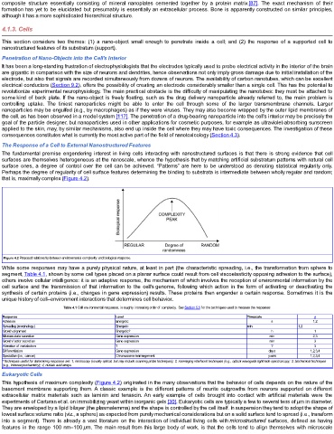
Perhaps the degree of regularity of cell surface features determining the binding to substrata is intermediate between wholly regular and random;
that is, maximally complex (Figure 4.2).
Figure 4.2 Proposed relationship between environmental complexity and biological response.
While some responses may have a purely physical nature, at least in part (the characteristic spreading, i.e., the transformation from sphere to
segment, Table 4.1, shown by some cell types placed on a planar surface could result from cell viscoelasticity opposing adhesion to the surface),
others involve cellular intelligence: it is an adaptive response, the mechanism of which involves the reception of environmental information by the
cell surface and the transmission of that information to the cell's genome, following which action in the form of activating or deactivating the
synthesis of certain proteins (i.e., changes in gene expression) results. These proteins then engender a certain response. Sometimes it is the
unique history of cell–environment interactions that determines cell behavior.
Table 4.1 Cell environmental responses, in roughly increasing order of complexity. See Section 5.5 for the techniques used to measure the responses
Response Level Timescale a
Adhesion Energetic s 1,2
Spreading (morphology) Energetic min 1,2
Growth alignment Energetic? h 1
Microexudate secretion Gene expression min 2,3
Growth factor secretion Gene expression min 3
Alteration of metabolism ? ? 3
Differentiation Gene expression days 1,2,3,4
Speciation (i.e., cancer) Chromosome rearrangement years 1,2,3,4
a Techniques useful for determning responses are: 1, mcroscopy (usually optical, but may include scanning probe techniques); 2, nonimaging interfacial techniques (e.g., optical waveguide lightmode spectroscopy; 3, biochemcal techniques
i
i
i
(e.g., immunocytochemstry); 4, nucleic acid arrays.
i
Eukaryotic Cells
This hypothesis of maximum complexity (Figure 4.2) originated in the many observations that the behavior of cells depends on the nature of the
basement membrane supporting them. A classic example is the different patterns of neurite outgrowths from neurons supported on different
extracellular matrix materials such as laminin and tenascin. An early example of cells brought into contact with artificial materials were the
experiments of Carturan et al. on immobilizing yeast within inorganic gels [30]. Eukaryotic cells are typically a few to several tens of μm in diameter.
They are enveloped by a lipid bilayer (the plasmalemma) and the shape is controlled by the cell itself. In suspension they tend to adopt the shape of
lowest surface:volume ratio (viz., a sphere) as expected from purely mechanical considerations but on a solid surface tend to spread (i.e., transform
into a segment). There is already a vast literature on the interaction of individual living cells with microstructured surfaces, defined as having
features in the range 100 nm–100 μm. The main result from this large body of work, is that the cells tend to align themselves with microscale