
Page 79 - Phase Space Optics Fundamentals and Applications
P. 79
60 Chapter Two
CCD
Rotation
axis
Insertion
device
0.001 - 1 m
Multilayer High resolution
monochromator Near field detector
Sample Fresnel diffraction
stage
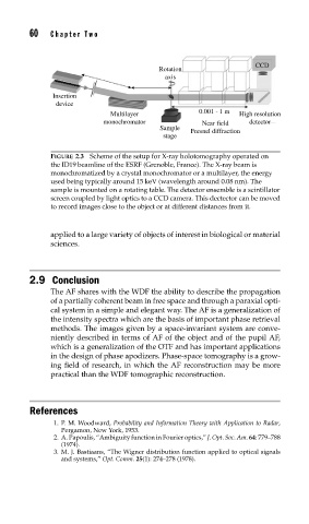
FIGURE 2.3 Scheme of the setup for X-ray holotomography operated on
the ID19 beamline of the ESRF (Grenoble, France). The X-ray beam is
monochromatized by a crystal monochromator or a multilayer, the energy
used being typically around 15 keV (wavelength around 0.08 nm). The
sample is mounted on a rotating table. The detector ensemble is a scintillator
screen coupled by light optics to a CCD camera. This dectector can be moved
to record images close to the object or at different distances from it.
applied to a large variety of objects of interest in biological or material
sciences.
2.9 Conclusion
The AF shares with the WDF the ability to describe the propagation
of a partially coherent beam in free space and through a paraxial opti-
cal system in a simple and elegant way. The AF is a generalization of
the intensity spectra which are the basis of important phase retrieval
methods. The images given by a space-invariant system are conve-
niently described in terms of AF of the object and of the pupil AF,
which is a generalization of the OTF and has important applications
in the design of phase apodizers. Phase-space tomography is a grow-
ing field of research, in which the AF reconstruction may be more
practical than the WDF tomographic reconstruction.
References
1. P. M. Woodward, Probability and Information Theory with Application to Radar,
Pergamon, New York, 1953.
2. A. Papoulis, “Ambiguity function in Fourier optics,” J. Opt. Soc. Am. 64: 779–788
(1974).
3. M. J. Bastiaans, “The Wigner distribution function applied to optical signals
and systems,” Opt. Comm. 25(1): 274–278 (1978).