Page 425 - Tandem Techniques
P. 425
Page 408

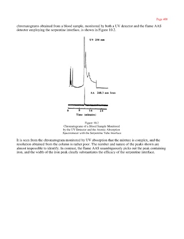
            chromatograms obtained from a blood sample, monitored by both a UV detector and the flame AAS
            detector employing the serpentine interface, is shown in Figure 10.2.


                                                         Figure 10.2
                                           Chromatograms of a Blood Sample Monitored
                                           by the UV Detector and the Atomic Absorption
                                           Spectrometer with the Serpentine Tube Interface

            It is seen from the chromatogram monitored by UV absorption that the mixture is complex, and the
            resolution obtained from the column is rather poor. The number and nature of the peaks shown are
            almost impossible to identify. In contrast, the flame AAS unambiguously picks out the peak containing
            iron, and the width of the iron peak clearly substantiates the efficacy of the serpentine interface.

