
Page 348 - Vibrational Spectroscopic Imaging for Biomedical Applications
P. 348
322 Cha pte r Ele v e n
11.2 The Birth of CARS Microscopy
11.2.1 First Generation CARS Microscopes
In microscopy, signals are collected from many spatially resolved loca-
tions in the sample, yielding images that typically consist of several
thousands to millions of pixels. Microscopic imaging is thus based on
the collection of many individual measurements, either sequentially
or in parallel. With such a large number of measurements, optical
microscopy relies on a contrast mechanism that is associated with a
high-photon flux. To build a microscope based on vibrational con-
trast, the CARS mechanism is a natural candidate, as the signal yields
are much higher than what the spontaneous Raman scattering pro-
cess can offer. The first Raman microscope was conceived in 1975, 22
but long image acquisition times had hampered the application of
this approach for imaging of dynamic samples such as live biological
specimens. In the early 1980s, Duncan et al. recognized the potential
advantage of CARS microscopy over the existing Raman microscope
in terms of imaging speed. In 1982, they constructed the first CARS
microscope. 23
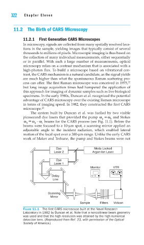
The system built by Duncan et al. was fuelled by two visible
picosecond dye lasers that provided the pump ω ω and Stokes
p
0
ω ω − ω beams for the CARS process (see Fig. 11.1). Before the
S 0 r
beams were focused to a 10-μm spot, a scanning mirror applied an
adjustable angle to the incident radiation, which enabled lateral
motion of the focal spot over a 300-μm range. Unlike the early CARS
work of Maker and Terhune, the pump and Stokes beams were not
Dye Mode Locked
Laser 2 Argon-Ion Laser
Dye
Laser 1
Monitor VTR
Delay
Line
Scanning
Mirror
Sample Filters Vidicon
FIGURE 11.1 The fi rst CARS microscope built at the Naval Research
Laboratory in 1982 by Duncan et al. Note that a noncollinear beam geometry
was used and that the high resolution was attained by the high numerical
detection lens. (Reproduced from Ref. 23, with permission of the Optical
Society of America.)