
Page 135 - Visions of the Future Chemistry and Life Science
P. 135
124 M. C. H. VAN DER MEULEN AND P. J. PRENDERGAST
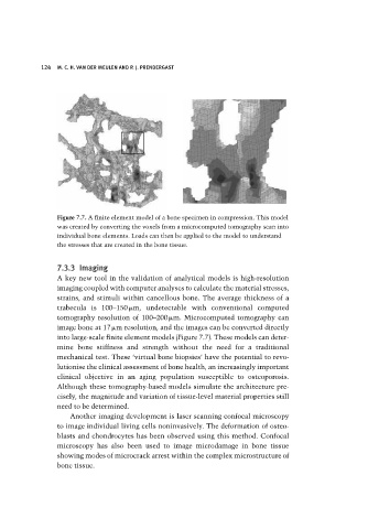
Figure 7.7. A finite element model of a bone specimen in compression. This model
was created by converting the voxels from a microcomputed tomography scan into
individual bone elements. Loads can then be applied to the model to understand
the stresses that are created in the bone tissue.
7.3.3 Imaging
A key new tool in the validation of analytical models is high-resolution
imaging coupled with computer analyses to calculate the material stresses,
strains, and stimuli within cancellous bone. The average thickness of a
trabecula is 100–150 m, undetectable with conventional computed
tomography resolution of 100–200 m. Microcomputed tomography can
image bone at 17 m resolution, and the images can be converted directly
into large-scale finite element models (Figure 7.7). These models can deter-
mine bone stiffness and strength without the need for a traditional
mechanical test. These ‘virtual bone biopsies’ have the potential to revo-
lutionise the clinical assessment of bone health, an increasingly important
clinical objective in an aging population susceptible to osteoporosis.
Although these tomography-based models simulate the architecture pre-
cisely, the magnitude and variation of tissue-level material properties still
need to be determined.
Another imaging development is laser scanning confocal microscopy
to image individual living cells noninvasively. The deformation of osteo-
blasts and chondrocytes has been observed using this method. Confocal
microscopy has also been used to image microdamage in bone tissue
showing modes of microcrack arrest within the complex microstructure of
bone tissue.