
Page 160 - Visions of the Future Chemistry and Life Science
P. 160
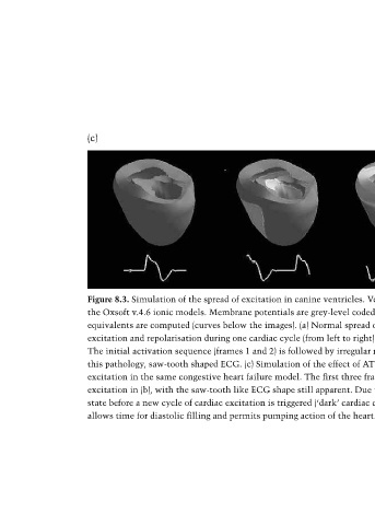
(c)


                 Figure 8.3. Simulation of the spread of excitation in canine ventricles. Ventricular cell models are based on a simplified version of
                 the Oxsoft v.4.6 ionic models. Membrane potentials are grey-level coded (dark – resting potential, light – action potential) and ECG
                 equivalents are computed (curves below the images). (a) Normal spread of excitation. Frames illustrate the normal sequence of
                 excitation and repolarisation during one cardiac cycle (from left to right). (b) Spread of excitation in a congestive heart failure model.
                 The initial activation sequence (frames 1 and 2) is followed by irregular re-entrant excitation (frames 3 and 4). Note the typical, for
                 this pathology, saw-tooth shaped ECG. (c) Simulation of the effect of ATP-modulated potassium channel openers on the spread of
                 excitation in the same congestive heart failure model. The first three frames are closely reminiscent of those leading to re-entrant
                 excitation in (b), with the saw-tooth like ECG shape still apparent. Due to the drug effect, however, the heart does reach a resting
                 state before a new cycle of cardiac excitation is triggered (‘dark’ cardiac chamber and ‘flat’ segment in the ECG, frame 4). This
                 allows time for diastolic filling and permits pumping action of the heart. From Kohl et al. 2000.