
Page 83 - Algae Anatomy, Biochemistry, and Biotechnology
P. 83
66 Algae: Anatomy, Biochemistry, and Biotechnology
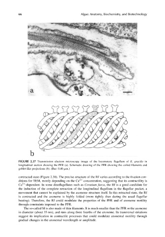
FIGURE 2.37 Transmission electron microscopy image of the locomotory flagellum of E. gracilis in
longitudinal section showing the PFR (a). Schematic drawing of the PFR showing the coiled filaments and
goblet-like projections (b). (Bar: 0.40 mm.)
contracted state (Figure 2.38). The precise structure of the Rf varies according to the fixation con-
ditions for TEM, mostly depending on the Ca 2þ concentration, suggesting that its contractility is
2þ
Ca -dependent. In some dinoflagellates such as Ceratium furca, the Rf is a good candidate for
the induction of the complete retraction of the longitudinal flagellum in the flagellar pocket, a
movement that cannot be explained by the axoneme structure itself. In this retracted state, the Rf
is contracted and the axoneme is highly folded (more tightly than during the usual flagellar
beating). Therefore, the Rf could modulate the properties of the PFR and of axoneme motility
through constraints imposed to the PFR.
The so-called Sf is also made of thin filaments. It is much smaller than the PFR or the axoneme
in diameter (about 35 nm), and runs along three fourths of the axoneme. Its transversal striations
suggest its implication in contractile processes that could modulate axonemal motility through
gradual changes in the axonemal wavelength or amplitude.