
Page 87 - Algae Anatomy, Biochemistry, and Biotechnology
P. 87
70 Algae: Anatomy, Biochemistry, and Biotechnology
FIGURE 2.42 Type 3 transition zone of Dinophyta (a), Glaucophyta (b), and Haptophyta (c).
the number of gyres, which in a short flagellum may be as low as one. This helix is present in Chry-
sophyceae, Xanthophyceae, and Eustigmatophyceae (Heterokontophyta).
Type 5 (Figure 2.45) is characterized by the so-called “stellate pattern”; typically, it is divided
into a longer distal and a shorter proximal part, separated by a basal plate. In longitudinal section,
the structure resembles an H, with the cross-bar located a short distance above the cell surface
(Figure 2.46). Transversely, the cross-bar may or may not extend to the peripheral doublets of
the axoneme. Variations regard the length of the proximal part, the location of the plate, and the
appearance of additional rings. This transition zone is typical of Chlorophyta.
Basal Bodies
A flagellum cannot be dissociated from its base, the basal body, or kinetosome. This structure has a
cylindrical form, with an average diameter of 0.2 mm and a variable height (average 0.5 mm). The
wall of the cylinder is discontinuous, and consists of nine microtubular triplets tilted to the radii at
an angle of 1308 and interconnected by transverse desmosomes. The complete tubule A consists of
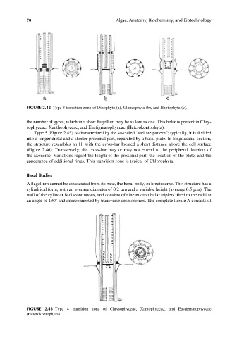
FIGURE 2.43 Type 4 transition zone of Chrysophyceae, Xantophyceae, and Eustigmatophyceae
(Heterokontophyta).