
Page 85 - Algae Anatomy, Biochemistry, and Biotechnology
P. 85
68 Algae: Anatomy, Biochemistry, and Biotechnology
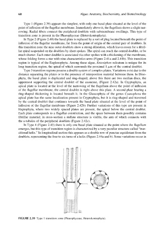
Type 1 (Figure 2.39) appears the simplest, with only one basal plate situated at the level of the
point of inflexion of the flagellar membrane. Immediately above it, the flagellum shows a slight nar-
rowing. Radial fibers connect the peripheral doublets with submembrane swellings. This type of
transition zone is present in the Phaeophyceae (Heterokontophyta).
In Type 2 (Figure 2.40) the basal plate is replaced by a sort of plug located beneath the point of
inflexion of the flagellar membrane, far from the point of origin of the central pair of doublets. In
this transition zone the nine outer doublets show a strong dilatation, which leaves room for a fibril-
lar spiral suspended on the doublets by short spokes. The spiral can reach the central doublet, or be
much shorter. Each outer doublet is associated via other spokes with a thickening of the membrane,
whose folding forms a star with nine characteristics arms (Figure 2.41a and 2.41b). This transition
region is typical of the Euglenophyta. Among these algae, Entosiphon sulcatum is unique for its
long transition region, the spiral of which surrounds the proximal 1 mm of the central doublet.
Type 3 transition regions possess a double system of complex plates. Variations exist due to the
distance separating the plates or to the presence of interposition material between them. In Dino-
phyta, the basal plate is duplicated and ring-shaped; above this there are two median discs, the
uppermost supporting the central doublet of the axoneme, (Figure 2.42a). In Cryptophyta, an
apical plate is located at the level of the narrowing of the flagellum above the point of inflexion
of the flagellar membrane; the central doublet is right above this plate. A second plate bearing a
ring-shaped thickening is located beneath it. In the Glaucophyta of the genus Cyanophora the
apical plate has the same localization present in Cryptophyta, but it is ring-shaped and traversed
by the central doublet that continues towards the basal plate situated at the level of the point of
inflexion of the flagellar membrane (Figure 2.42b). Further variations of this type are present in
Haptophyta, where two widely spaced plates are present, the apical below the central doublet.
Each plate corresponds to a flagellar constriction, and the space between them possibly contains
fibrillar material; in cross-section a stellate structure is visible, the arm of which connects with
the a-tubules of the peripheral doublets (Figure 2.42c).
In Type 4 (Figure 2.43) there is only one basal plate situated at the point where the flagellum
emerges, but this type of transition region is characterized by a very peculiar structure called “tran-
sitional helix.” In longitudinal section this appears as a double row of punctae equidistant from the
doublets, representing the four to six turns of a helix (Figure 2.44a and b). Some variations occur in
FIGURE 2.39 Type 1 transition zone (Phaeophyceae, Heterokontophyta).