
Page 25 - A Practical Introduction to Optical Mineralogy
P. 25
THE MICROSCOPIC STUDY OF MINERALS THE REFLECTED-LIGHT MICROSCOPE
Zoning is generally a growth phenomenon and is therefore related to
the crystal shape.
Dispersion
Refractive index increases as the wavelength oflight decreases. Thus the
refractive index of a mineral for red light is less than for blue light (since
the wavelength of red light is greater than the wavelength of blue light).
White light entering a mineral section is split into the colours of the
spectrum, with blue nearest to the normal (i.e. the straight through path)
and red the furthest away. This breaking up of the white light is called
dispersion. In most minerals the amount of dispersion is very small and
will not affect the mineral's optical properties. However, the Na-rich
l~>,-----4- heat absorbing filter
clinopyroxenes, the Na-rich amphiboles, sphene, chloritoid, zircon and aperture diaphragm
?rookite possess very strong dispersion. With many of these minerals, Smith
illuminator field
mterference figures may be difficult to obtain and the use of accessory .,__ ______ diaphragm
plates (to determine mineral sign etc.) may not be possible.
l.~~:::l-..b;==------- focusing
revolving control
Each mineral possesses a few diagnostic properties, and in the descrip- objective
changer
tions in Chapter 2 these have been marked with an asterisk. Sometimes
a final paragraph discusses differences between the mineral being stage
centring ------<1----1--"'""
described and other minerals that have similar optical properties. screws
coarse
focus
1.4 The reflected-light microscope
The light source fine focus
A high intensity source (Fig. 1.3) is required for reflected-light studies,
mainly because of the low brightness of crossed polar images.
Tungsten-halogen quartz lamps are used, similar to those in transpa-
rency projectors, and the tungsten light (A source) gives the field a
yellowish tint. Many microscopists prefer to use a blue correction filter
to change the light colour to that of daylight (C source). A monochro-
matic light source (coloured light corresponding to a very limited range
of the visible spectrum) is rarely used in qualitative microscopy, but
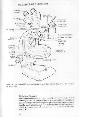
Figure 1.3 The Vickers M73 reflected light microscope. Note that it is the polariser that rotates in
monochromatic filters for the four standard wavelengths ( 4 70 nm,
!his microscope.
546 nm, 589 nm and 650 nm) could be useful in comparing the
brightness of coexisting minerals, especially now that quantitative
measurements of brightness are readily available.
The polariser The incident illuminator
Polarised light is usually obtained by using a polarising film, and this The incident illuminator sits above the objective and its purpose is to
should be protected from the heat of the lamp by a glass heat filter. The reflect light down through the objective on to the polished specimen. As
polariser should always be inserted in the optical train. It is best fixed in the reflected light travels back up through the objective to the eyepiece it
orientation to give E-W vibrating incident light. However, it is useful to must be possible for this light to pass through the incident illuminator.
be able to rotate the polariser on occasion in order to correct its orien- There are three types of reflector used in incident illuminators
(Fig. 1.4):
tation or as an alternative to rotating the analyser.
12 13