
Page 274 - Advances in Biomechanics and Tissue Regeneration
P. 274
272 14. USING 3-D PRINTING AND BIOPRINTING TECHNOLOGIES FOR PERSONALIZED IMPLANTS
In the indirect bioprinting approach, first, a negative supportive mold is created, Fig. 14.1D. This approach is widely
used for the fabrication of vasculatures in which a sacrificial ink is easily removed, where this fugitive ink was used to
print tubular networks within the construct [3]. One example is a recent research developed by Kang et al. that they
describe a system for deposition of cell-laden hydrogels together with synthetic biodegradable polymers in integrated
patterns and anchored on sacrificial hydrogels [20]. The obtained cell-laden hydrogel is important to protect cell via-
bility and to promote growth and expansion; at the same time, the adjacent sacrificial scaffolding (Pluronic F127) was
used to provide the initial structural and architectural integrity.
The direct bioprinting approach can also be used as an alternative for organ fabrication. Bertassoni et al. [21] used
the direct bioprinting to precisely deposit cells and cell-laden materials with the objective of generating controlled tis-
sue architecture [22]. Their work shows a strategy for bioprinting of photolabile cell-laden methacrylated gelatin
(GelMA) hydrogels in which encapsulated hepatocyte cells preserved high cell viability for at least 8days.
14.3 MATERIALS
Depending on the printing technique, the composition of the materials used in 3-D bioprinting processes will differ.
It is, therefore, necessary to define the differences of the materials used according to the different printing techniques to
determine the improvement that each compound brings to the print quality [23]. Those materials are known as
“bioinks,” which is used as a term making reference to original conventional inkjet printing inks, means the bioprin-
table materials used in 3-D bioprinting processes in which cells are deposited in a spatially controlled pattern to
fabricate living tissues and organs. In this section, properties of materials suitable as bioinks, particularly
hydrogel-forming materials, used in laser-, inkjet- and extrusion-based bioprinting will be described.
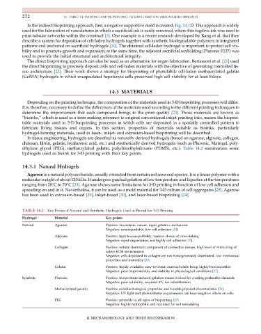
In tissue engineering, hydrogels are classified as naturally derived hydrogels (based on agarose, alginate, collagen,
chitosan, fibrin, gelatin, hyaluronic acid, etc.) and synthetically derived hydrogels (such as Pluronic, Matrigel, poly-
ethylene glycol (PEG), methacrylated gelatin, polydimethylsiloxane (PDMS), etc.). Table 14.2 summarizes some
hydrogels used as bioink for 3-D printing with their key points.
14.3.1 Natural Hydrogels
Agarose is a natural polysaccharide, usually extracted from certain red seaweed species. It is a linear polymer with a
molecular weight of about 120kDa. It undergoes gradual gelation at low temperature and liquefies at the temperatures
ranging from 20°Cto70°C [23]. Agarose shows some limitations for 3-D printing in function of low cell adhesion and
spreading on and in it. Nevertheless, it can be used as a mold material for 3-D culture of cell aggregates [28]. Agarose
has been used in extrusion-based [29], inkjet-based [30], and laser-based bioprinting [24].
TABLE 14.2 Key Points of Natural and Synthetic Hydrogels Used as Bioink for 3-D Printing
Hydrogel Material Key points
Natural Agarose Positive: viscoelastic nature, rapid gelation mechanism
Negative: nondegradable, low cell adhesion [24]
Alginate Positive: high biocompatibility, various choice of cross-linking
Negative: rapid degradation, not highly cell adhesive [25]
Collagen Positive: natural dominant component of connective tissues, high level of mimicking of
native ECM environment.
Negative: cells deposited in collagen are not homogeneously distributed, low mechanical
properties and instability [23]
Gelatin Positive: highly available, easy-to-obtain material while being highly biocompatible
Negative: poor bioprintability and stability in physiological conditions [17]
Synthetic Pluronic Positive: temperature-induced gelation makes it ideal for creating perfusable channels
Negative: poor solubility, required 4°C for solubilization
Methacrylated gelatin Positive: suitable biological properties and tunable physical characteristics [26]
Negative: UV light and photoinitiator requirements can have negative effects on cells
PEG Positive: printable in all types of bioprinting [27]
Negative: highly hydrophilic and not ideal for cell remodeling
II. MECHANOBIOLOGY AND TISSUE REGENERATION