
Page 273 - Advances in Biomechanics and Tissue Regeneration
P. 273
14.2 BIOPRINTING 271
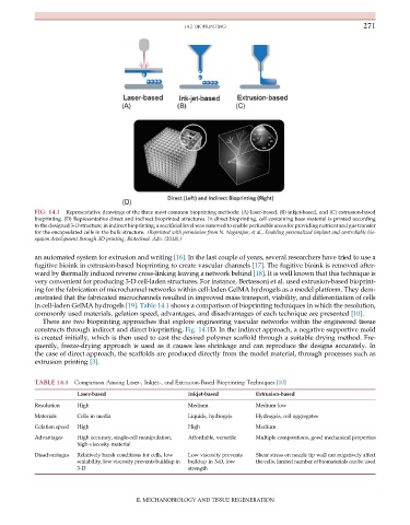
FIG. 14.1 Representative drawings of the three most common bioprinting methods: (A) laser-based, (B) inkjet-based, and (C) extrusion-based
bioprinting. (D) Representative direct and indirect bioprinted structures. In direct bioprinting, cell containing base material is printed according
to the designed 3-D structure; in indirect bioprinting, a sacrificial level was removed to enable perfusable areas for providing nutrient and gas transfer
for the encapsulated cells in the bulk structure. (Reprinted with permission from N. Nagarajan, et al., Enabling personalized implant and controllable bio-
system development through 3D printing, Biotechnol. Adv. (2018).)
an automated system for extrusion and writing [16]. In the last couple of years, several researchers have tried to use a
fugitive bioink in extrusion-based bioprinting to create vascular channels [17]. The fugitive bioink is removed after-
ward by thermally induced reverse cross-linking leaving a network behind [18]. It is well known that this technique is
very convenient for producing 3-D cell-laden structures. For instance, Bertassoni et al. used extrusion-based bioprint-
ing for the fabrication of microchannel networks within cell-laden GelMA hydrogels as a model platform. They dem-
onstrated that the fabricated microchannels resulted in improved mass transport, viability, and differentiation of cells
in cell-laden GelMA hydrogels [19]. Table 14.1 shows a comparison of bioprinting techniques in which the resolution,
commonly used materials, gelation speed, advantages, and disadvantages of each technique are presented [10].
There are two bioprinting approaches that explore engineering vascular networks within the engineered tissue
constructs through indirect and direct bioprinting, Fig. 14.1D. In the indirect approach, a negative supportive mold
is created initially, which is then used to cast the desired polymer scaffold through a suitable drying method. Fre-
quently, freeze-drying approach is used as it causes less shrinkage and can reproduce the designs accurately. In
the case of direct approach, the scaffolds are produced directly from the model material, through processes such as
extrusion printing [3].
TABLE 14.1 Comparison Among Laser-, Inkjet-, and Extrusion-Based Bioprinting Techniques [10]
Laser-based Inkjet-based Extrusion-based
Resolution High Medium Medium-low
Materials Cells in media Liquids, hydrogels Hydrogels, cell aggregates
Gelation speed High High Medium
Advantages High accuracy, single-cell manipulation, Affordable, versatile Multiple compositions, good mechanical properties
high-viscosity material
Disadvantages Relatively harsh conditions for cells, low Low viscosity prevents Shear stress on nozzle tip wall can negatively affect
scalability, low viscosity prevents buildup in buildup in 3-D, low the cells, limited number of biomaterials can be used
3-D strength
II. MECHANOBIOLOGY AND TISSUE REGENERATION