
Page 280 - Advances in Biomechanics and Tissue Regeneration
P. 280
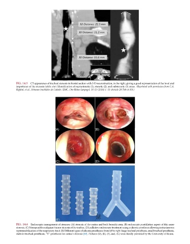
FIG. 14.5  CT appearance of tracheal stenosis in frontal section with 3-D reconstruction, in the right, giving a good representation of the level and
           importance of the stenosis (white star). Identification of suprastenotic (1), stenotic (2), and substenotic (3) areas. (Reprinted with permission from C.A.
           Righini, et al., St  enoses trach  eales de l’adulte. EMC, Oto-Rhino-Laryngol. 10 (1) (2014) 1–15 (Article 20-760-A-10).)


           FIG. 14.6  Endoscopic management of stenoses: (A) stenosis of the carina and both bronchi stem, (B) endoscopic postdilation aspect of this same
           stenosis, (C) Nonoperable malignant tumor stenosis of the trachea, (D) palliative endoscopic treatment using a silicone prosthesis allowing instantaneous
           repermeabilization of the respiratory tract. (E) Different types of silicone prostheses: from left to right: large tracheal prosthesis, small tracheal prosthesis,
           diabolo tracheal prosthesis, “Y” prosthesis for carina’s stenosis [68]. Pictures (D), (E), (F), and, (G) were kindly provided by the University of Reims.