
Page 372 - Advances in Biomechanics and Tissue Regeneration
P. 372
18.4 ARTICULAR CARTILAGE AND TISSUE ENGINEERING 369
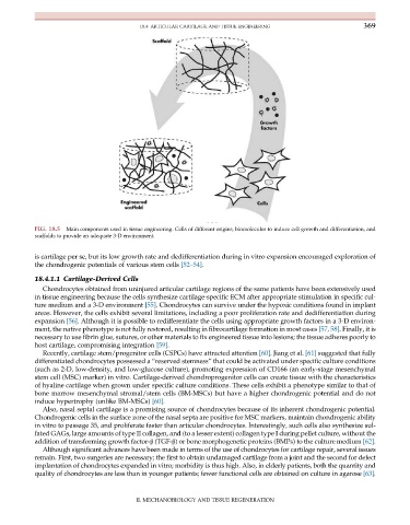
FIG. 18.5 Main components used in tissue engineering. Cells of different origins, biomolecules to induce cell growth and differentiation, and
scaffolds to provide an adequate 3-D environment.
is cartilage per se, but its low growth rate and dedifferentiation during in vitro expansion encouraged exploration of
the chondrogenic potentials of various stem cells [52–54].
18.4.1.1 Cartilage-Derived Cells
Chondrocytes obtained from uninjured articular cartilage regions of the same patients have been extensively used
in tissue engineering because the cells synthesize cartilage-specific ECM after appropriate stimulation in specific cul-
ture medium and a 3-D environment [55]. Chondrocytes can survive under the hypoxic conditions found in implant
areas. However, the cells exhibit several limitations, including a poor proliferation rate and dedifferentiation during
expansion [56]. Although it is possible to redifferentiate the cells using appropriate growth factors in a 3-D environ-
ment, the native phenotype is not fully restored, resulting in fibrocartilage formation in most cases [57, 58]. Finally, it is
necessary to use fibrin glue, sutures, or other materials to fix engineered tissue into lesions; the tissue adheres poorly to
host cartilage, compromising integration [59].
Recently, cartilage stem/progenitor cells (CSPCs) have attracted attention [60]. Jiang et al. [61] suggested that fully
differentiated chondrocytes possessed a “reserved stemness” that could be activated under specific culture conditions
(such as 2-D, low-density, and low-glucose culture), promoting expression of CD166 (an early-stage mesenchymal
stem cell (MSC) marker) in vitro. Cartilage-derived chondroprogenitor cells can create tissue with the characteristics
of hyaline cartilage when grown under specific culture conditions. These cells exhibit a phenotype similar to that of
bone marrow mesenchymal stromal/stem cells (BM-MSCs) but have a higher chondrogenic potential and do not
induce hypertrophy (unlike BM-MSCs) [60].
Also, nasal septal cartilage is a promising source of chondrocytes because of its inherent chondrogenic potential.
Chondrogenic cells in the surface zone of the nasal septa are positive for MSC markers, maintain chondrogenic ability
in vitro to passage 35, and proliferate faster than articular chondrocytes. Interestingly, such cells also synthesize sul-
fated GAGs, large amounts of type II collagen, and (to a lesser extent) collagen type I during pellet culture, without the
addition of transforming growth factor-β (TGF-β) or bone morphogenetic proteins (BMPs) to the culture medium [62].
Although significant advances have been made in terms of the use of chondrocytes for cartilage repair, several issues
remain. First, two surgeries are necessary; the first to obtain undamaged cartilage from a joint and the second for defect
implantation of chondrocytes expanded in vitro; morbidity is thus high. Also, in elderly patients, both the quantity and
quality of chondrocytes are less than in younger patients; fewer functional cells are obtained on culture in agarose [63].
II. MECHANOBIOLOGY AND TISSUE REGENERATION