
Page 367 - Advances in Biomechanics and Tissue Regeneration
P. 367
364 18. CARTILAGE REGENERATION AND TISSUE ENGINEERING
contents correlate strongly with the cartilage creep modulus, indicating that compressive stiffness is determined by the
GAGs rather than collagen [12]. Other proteoglycans of the chondral matrix are biglycan, decorin, fibromodulin, and
perlecan [13]. Proteoglycans of various types associate to form larger ECM polymeric complexes termed aggregates;
about 300 proteoglycan molecules attach to a linear molecule of HA (a huge GAG containing repeats of up to 25,000
nonsulfated disaccharide units) via linkage proteins, forming the large proteoglycan aggregates mentioned earlier.
Sulfated aggrecan GAGs are highly negatively charged and therefore strongly hydrophilic, forming highly
hydrated gels; in fact, water represents 70%–80% of cartilage net weight. Water does not flow out of articular cartilage
when walking [6], but transient changes in water content do occur during joint movement and when joints are sub-
jected to pressure; this allows, on the one hand, rapid diffusion of water-soluble molecules from blood vessels in sur-
rounding tissues toward chondrocytes and, on the other hand, creation of a swelling pressure or turgor allowing the
ECM to withstand compressive forces. The low-friction surface explains ECM resilience, and the 3-D meshwork of
tension-resisting collagen fibrils allows the swelling pressure imparted by proteoglycans to be withstood; cartilage
is well adapted to high intermittent pressures and efficiently resists shear stress imposed on synovial joints [14].
Glycoproteins are adhesive proteins featuring a short, branched oligosaccharide chain with multiple domains, each
of which specifically binds other macromolecules of the ECM or receptors on the cell surface. These glycoproteins are
important in terms of cellular interactions, organizing and assembling other ECM components, and helping cells attach
to the ECM.
Within the chondral ECM, various glycoproteins such as laminin or fibronectin are detected at different concentra-
tions during the life span and in periods of good health or illness [13]. The glycoprotein tenascin is abundant during
cartilage development, but the level decreases during maturation; this glycoprotein disappears almost completely in
adult articular cartilage, although it has been detected in both the cartilage and synovium of osteoarthritis patients [15].
The glycoproteins lubricin and chitinase 3-like-1 (CHI3L1) are present in normal articular cartilage, produced by
both articular chondrocytes and other cell types. Lubricin is abundant in normal cartilage but decreases in patients
with osteoarthritis; this is one of the major joint lubricants and is considered to be chondroprotective, preventing car-
tilage wear and reducing the amount of friction on the surface of articular cartilage [3]. However, glycoprotein CHI3L1
has the opposite effect, being associated with mediators of inflammation and cartilage damage during the pathogen-
esis of osteoarthritis [16].
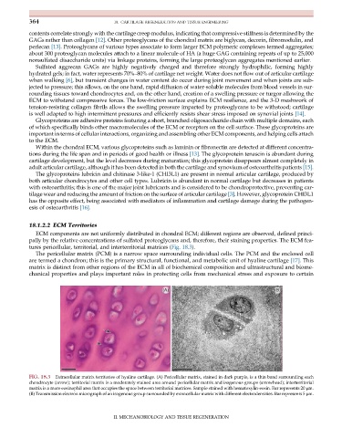
18.1.2.2 ECM Territories
ECM components are not uniformly distributed in chondral ECM; different regions are observed, defined princi-
pally by the relative concentrations of sulfated proteoglycans and, therefore, their staining properties. The ECM fea-
tures pericellular, territorial, and interterritorial matrices (Fig. 18.3).
The pericellular matrix (PCM) is a narrow space surrounding individual cells. The PCM and the enclosed cell
are termed a chondron; this is the primary structural, functional, and metabolic unit of hyaline cartilage [17].This
matrix is distinct from other regions of the ECM in all of biochemical composition and ultrastructural and biome-
chanical properties and plays important roles in protecting cells from mechanical stress and exposure to certain
FIG. 18.3 Extracellular matrix territories of hyaline cartilage. (A) Pericellular matrix, stained in dark purple, is a thin band surrounding each
chondrocyte (arrow); territorial matrix is a moderately stained area around pericellular matrix and isogenous groups (arrowhead); interterritorial
matrix is a more eosinophil area that occupies the space between territorial matrices. Sample stained with hematoxylin-eosin. Bar represents 20 μm.
(B) Transmission electron micrograph of an isogenous group surrounded by extracellular matrix with different electrodensities. Bar represents 5 μm.
II. MECHANOBIOLOGY AND TISSUE REGENERATION