
Page 366 - Advances in Biomechanics and Tissue Regeneration
P. 366
18.1 CARTILAGE TISSUE 363
processes. However, Hunziker et al. [6] showed that when cartilage is processed using specific methods, such as
high-pressure freezing, freeze-substitution, and low-temperature embedding, the chondrocytes remain in an
expanded state and empty spaces around cells are lacking.
3
7
The chondrocyte density is 1.4 10 cells/cm in mature cartilage [7], indicating that cells occupy less than 5% of the
total cartilage volume but are essential for ECM production and maintenance throughout life. The cells are metabol-
ically active; have high nutritional requirements; and, although suited to low-oxygen environments because of the
absence of blood vessels, in fact, consume oxygen (although at lower rates than other cell types) and are susceptible
to damage caused by oxidative stress [8, 9].
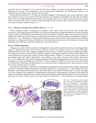
18.1.2 Hyaline Cartilage Extracellular Matrix [1, 2, 10]
Hyaline cartilage exhibits a homogeneous amorphous matrix when stained and observed with standard light
microscopy. ECM components are secreted locally and then assembled into an organized meshwork closely associated
with the surfaces of chondroblasts and chondrocytes that produce them. The ECM contains many components that can
modify matrix behavior; the three major macromolecules are the fibrous proteins collagen, proteoglycans, and glyco-
proteins (Fig. 18.2), embedded in a highly hydrated gel-like ground substance that confers physical and functional
properties to cartilage and, therefore, the ability to resist compressive forces because of the strength of collagen while
permitting rapid diffusion of nutrients, metabolites, and waste products.
18.1.2.1 ECM Components
Collagens are a family of fibrous proteins including fibril-forming, fibril-associated, and network-forming proteins,
present in different proportions in the diverse connective tissues. The collagen molecule has a long, stiff, triple-
stranded helical structure; three collagen polypeptide chains (α-chains) extremely rich in proline and glycine residues
are wrapped around one another to form a ropelike superhelix that is highly resistant to tension. These collagen mol-
ecules assemble into higher-order polymers termed collagen fibrils (10–300 nm in diameter and many hundreds of
micrometers long) that exhibit characteristic cross striations every 68 nm observed with electron microscopy
(Fig. 18.2C), attributable to the regularly staggered packing of individual collagen molecules. In hyaline cartilage, type
II collagen, found only in cartilage, accounts for 90%–95% of all collagens in the ECM [3] and forms a three-dimensional
(3-D) meshwork of fibrils that are thinner than those of the typical type I collagen present in other connective tissues
that contains different types of α-chains.
Proteoglycan monomers are formed from abundant glycosaminoglycan (GAG) chains; these are linear unbranched
polymers of repeating sulfated disaccharides covalently attached to a core protein (Fig. 18.2B). Sulfated GAGs are
basophilic when stained with hematoxylin or other basic dyes and also exhibit metachromasia. The major proteogly-
can of the cartilage ECM is aggrecan, a cartilage-specific proteoglycan. Each aggrecan monomer contains about 100
chains of chondroitin sulfate and 60 of keratan sulfate, featuring three globular domains that bind noncovalently to
hyaluronic acid (HA), matrix proteins, and other unknown substances [11]. The chondroitin and keratan sulfate
FIG. 18.2 Chondral extracellular matrix.
(A) Chondrocytes surrounded by collagen
fibrils and aggregates of proteoglycans.
(B) Proteoglycan monomer with abundant
glycosaminoglycan chains attached to a core
protein, which is bound to hyaluronic acid by
means of link proteins. (C) Enlarged detail
showing transmission electron micrograph
of the extracellular matrix, where type II col-
lagen fibrils can be identified (arrows). Bar
represents 500 nm.
II. MECHANOBIOLOGY AND TISSUE REGENERATION