
Page 368 - Advances in Biomechanics and Tissue Regeneration
P. 368
18.1 CARTILAGE TISSUE 365
molecules, ensuring that articular cartilage is not degraded [13, 18]. The PCM stains densely with basic dyes
because it contains high concentrations of sulfated GAGs and glycoproteins, but only some loose collagen fibrils,
creating a meshlike capsular ultrastructure that modifies the mechanical load imparted to the chondrocyte; in fact,
the elastic modulus is significantly lower than that of the bulk ECM [17, 18]. Aggrecan is anchored to both cell
surfaces and HA, and this is of particular importance within the PCM; Knudson et al. [19] concluded that both
aggrecan and HA contribute to the establishment and maintenance of cell-cell spaces between chondrocytes.
PCM contains type VI and IX collagens but lacks type II collagen; thus, under normal conditions, type II collagen
fibrils are not exposed to chondrocytes [13]. PCM also contains perlecan, a large proteoglycan unique to the PCM,
which colocalizes with type VI collagen [13].
The second ECM territory is the territorial matrix that surrounds the PCM and isogenous groups; this matrix stains
moderately. The matrix contains abundant type II collagen fibrils that are irregularly distributed and GAGs and pro-
teoglycans (but at lower concentrations than the PCM).
Finally, the interterritorial matrix occupies the space between territorial matrices. This matrix stains slightly with
basic dyes and contains large amounts of type II collagen fibrils, glycoproteins, and aggrecan, associated with HA (as
in other territories), thus supporting biomechanical function [19].
18.1.3 Synovial Joints
Diarthroses are bone articulations that permit wide joint movements. The joints have capsules that join both bone
ends. The capsules have two layers; the outermost is formed from dense connective tissue containing blood vessels and
nerves in continuity with the periosteum of the bones joined at the edge of the articular cartilage. The inner layer is a
synovial membrane that covers the synovial or articular cavity. The inner surface of the synovial membrane is lined
with one or two layers of synovial cells covering a loose connective tissue with abundant fenestrated capillaries. There
are two types of synovial cells: type A synoviocytes are macrophage-like, and type B synoviocytes are fibroblast-like.
The articular cavity contains the synovial fluid, a liquid that reduces friction between the hyaline cartilages covering
both articular surfaces. Synovial liquid is produced by the synovial cells and is an ultrafiltrated blood plasma contain-
ing abundant HA (which serves as a lubricant) [10]. The liquid contains leukocytes and glycoproteins such as lubricin
(secreted into the synovial cavity by both type B synoviocytes and superficial chondrocytes); this glycoprotein reduces
friction within the joint [13].
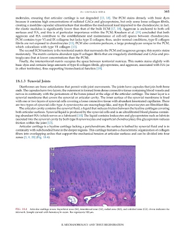
Articular cartilage is a hyaline cartilage lacking a perichondrium; the surface is bathed by synovial fluid and is in
continuity with subchondral bone in the deeper regions. This cartilage features a characteristic organization of collagen
fibers into overlapping arches that support the mechanical tension at articular surfaces and can be divided into four
zones [3, 9, 20] (Fig. 18.4):
FIG. 18.4 Articular cartilage zones. Superficial zone (SZ), transitional zone (TZ), radial zone (RZ), and calcified zone (CZ). Arrow indicates the
tidemark. Sample stained with hematoxylin-eosin. Bar represents 100 μm.
II. MECHANOBIOLOGY AND TISSUE REGENERATION