
Page 193 - An Introduction to Microelectromechanical Systems Engineering
P. 193
172 MEMS Applications in Life Sciences
section, flow is laminar: the fluid can be envisioned as flowing in laminar sheets,
moving slowest at the edges due to the drag of the walls and moving fastest at the
center. For higher Reynolds numbers, the flow is turbulent rather than laminar. In
3
microfluidics, water-based solutions are usually used, having ρ 1 g/cm and µ 0.01
g/(cm•s). For a representative hydraulic diameter of 30 µm and a representative
velocity of 1 mm/s, the Reynolds number is merely 0.03. In microfluidics, Reynolds
numbers are usually below one [4].
This has great implications for mixing in microfluidics. In the macroscopic
world, simply joining two channels together would enable the two streams to inter-
mix. At these low Reynolds numbers, however, streams joined from two channels
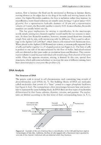
simply flow side by side, with intermixing only by diffusion. This is used to advan-
tage in the Agilent Cell LabChip , which detects cells stained with fluorescent dyes.
When placed in the Agilent 2100 Bioanalyzer system, a vacuum pulls separate flows
of cells and buffer together in a Y-shaped junction (see Figure 6.2). The flow of cells
is pushed to one side of the microchannel by the flow of buffer. Individual stained
cells are detected as they pass under an excitation beam and fluoresce. This concen-
tration scheme is used because individual cells would clog a flow channel of the same
width. Often the opposite situation, mixing, is desired. In this case, special flow
structures, which add some turbulence or increase the area of diffusive mixing, have
been demonstrated to overcome this problem [5].
DNA Analysis
The Structure of DNA
The genetic code is stored in cell chromosomes, each containing long strands of
deoxyribonucleic acid (DNA) [6, 7]. The building blocks of DNA are molecules
called nucleotides that consist of a “base” joined to a sugar-phosphate backbone
[see Figure 6.3(a)]. The nomenclature often interchanges between base and nucleo-
tide to represent the same building block. In DNA there are four types of nucleotides
differentiated by their bases: adenine, thymine, cytosine, and guanine. The nucleo-
tides are labeled according to the first letter of their corresponding bases: A, T, C,
Cell stained with Focused Fluorescence
fluorescent dye excitation beam
Stained cells F F F F F F Vacuum
F
Buffer
Figure 6.2 Example of the use of laminar flow in microfluidics: In the Cell LabChip from Agilent
Technologies of Palo Alto, California, the flow of cells tagged with a fluorescent dye is pushed to
one side of the channel. Individual cells are detected when they fluoresce.