Page 198 - An Introduction to Microelectromechanical Systems Engineering
P. 198
DNA Analysis                                                                  177

                                                      V


                      Starting point  Electric field  Negative ion (DNA) motion  Smaller DNA fragments
                                                                      travel farther, spread more
                                                     (a)

                      Reference                         Reference

                       Unknown                          Unknown
                               Unknown matches reference     Unknown does not match reference
                                                     (b)


                       Original copy = ....ATCGCTAGTCAGAT....
                       Solution with stop-at-C
                                                   ....ATCGCTAGTCAGAT....
                                                    TAGC  stop
                                           ....ATCGCTAGTCAGAT....
                                                              ....ATCGCTAGTCAGAT....
                                            TAGCGATC  stop
                                                                TAGCGATCAGTC  stop
                                                     (c)

                                       400          300          200          100
                                       bases        bases        bases        bases
                                       long         long         long         long
                       Stop at C
                       Stop at G
                       Stop at A
                       Stop at T
                                                     (d)
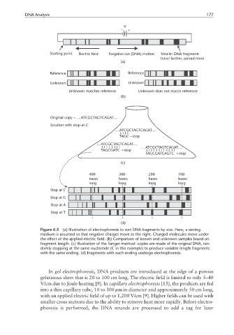
                  Figure 6.5  (a) Illustration of electrophoresis to sort DNA fragments by size. Here, a sieving
                  medium is assumed so that negative charges move to the right. Charged molecules move under
                  the effect of the applied electric field. (b) Comparison of known and unknown samples based on
                  fragment length. (c) Illustration of the Sanger method: copies are made of the original DNA, ran-
                  domly stopping at the same nucleotide (C in this example) to produce variable-length fragments
                  with the same ending. (d) Fragments with each ending undergo electrophoresis.


                      In gel electrophoresis, DNA products are introduced at the edge of a porous
                  gelatinous sheet that is 20 to 100 cm long. The electric field is limited to only 5–40
                  V/cm due to Joule heating [9]. In capillary electrophoresis [15], the products are fed
                  into a thin capillary tube, 10 to 300 µm in diameter and approximately 50 cm long,
                  with an applied electric field of up to 1,200 V/cm [9]. Higher fields can be used with
                  smaller cross sections due to the ability to remove heat more rapidly. Before electro-
                  phoresis is performed, the DNA strands are processed to add a tag for later

