
Page 199 - An Introduction to Microelectromechanical Systems Engineering
P. 199
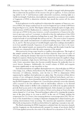
178                                                  MEMS Applications in Life Sciences

                                                       32
                 detection. One type of tag is radioactive ( P), which is imaged with photographic
                 film to determine the position of the strand in the gel or capillary. A more common
                 tag added to the 5’ end fluoresces under ultraviolet excitation, emitting light at a
                 visible wavelength. Used alone, electrophoretic separation can compare two samples
                 of fragments of DNA to determine whether they match but cannot tell the exact
                 sequence.
                    If electrophoresis is to be employed to determine the sequence of bases on a sec-
                 tion of DNA, the Sanger method may be used for fragments up to about 1,000 bases
                 long [see Figure 6.5(c, d)] [9]. This begins with many identical copies of single, dena-
                 tured sections of DNA. Replication in a solution with dNTPs is started from the 5’
                 end, just as in PCR. In this case, however, a small concentration of bases in the solu-
                 tion of one type, such as C (cytosine), is altered so that the replication of that DNA
                 strand stops when the replication-halting base is used. This results in copies of the
                 original strands of varying length that always end in C. The same is done in separate
                 solutions with small concentrations of replication-halting bases of the other types
                 (G, A, and T). The four groups of variable-length copies then undergo electrophore-
                 sis in four parallel channels. Sequences of each length, from one base to the maxi-
                 mum in the original sample, are separated for reading, and the results from the four
                 channels are compared to infer the entire sequence of the strand.
                    Miniaturization brings many benefits to capillary electrophoresis. The length of
                 the sample emitted into the channel can be kept relatively short (on the order of
                 100 µm), reducing the distance that must be traveled for the fragments of different
                 lengths to separate. Reducing the length of the channel decreases the applied voltage
                 required to maintain a high electric field from a few kilovolts down to hundreds of
                 volts. Faster separation times also become possible because the molecules have to
                 travel shorter distances. Additionally, the overall volume of DNA and reagents
                 decreases significantly to one microliter or less.
                    Early demonstrations of capillary electrophoresis on a chip took place in 1992 at
                 Ciba-Geigy, Ltd., of Basle, Switzerland [16]. Woolley and Mathies [17, 18] from the
                 University of California, Berkeley, were the first in 1994 to demonstrate DNA
                 sequencing by capillary electrophoresis on a glass chip. The structure of their device
                 consists of two orthogonal channels etched with buffered hydrofluoric acid into a
                 first glass substrate: a short channel for injecting fluid and a long channel for separat-
                 ing the DNA fragments (see Figure 6.6). A second glass substrate covers the channels
                 and is secured to the first substrate with an intermediate adhesive or by thermal
                 bonding. Holes etched or drilled with a diamond-core drill in the top glass substrate
                 provide fluid access ports to the embedded channels. Both channels are typically 50
                 µm wide and 8 µm deep but can be as wide as 100 µm and as deep as 16 µm; the sepa-
                 ration channel is 3.5 cm long. Thermal bonding is achieved by ramping the tempera-
                 ture of the glass plates in an oven to 600°C at the rate of 5°C/min, holding the
                 temperature for 2 to 3 hours, then ramping down to room temperature [18]. The sur-
                 faces of the channels have a coating to eliminate charging due to deprotonation, pre-
                 venting electroosmosis from occurring. The injection and separation channels are
                 filled with sieving matrix of hydroxyethylcellulose by applying a vacuum to one end.
                    The fluid containing the DNA fragments is admitted into the injection channel,
                 and the fragments are electrophoretically pumped by means of an electric field of
                 170 V/cm applied across the two ends of the channel for a duration of 30–60s. The