
Page 202 - An Introduction to Microelectromechanical Systems Engineering
P. 202
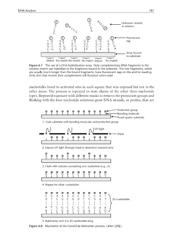
DNA Analysis                                                                  181

                                                      F
                                  F                   G    A G           Unknown strands
                                  A      F            C      C
                                 T        A T          G     G F         in solution
                                 C           C G       A
                                 G
                               F      F      F       F      F      F        Fluorescent
                                                           -
                             T-A    A A     T A    G G    CG     C G        tag
                             A-T    A T     A T    G C    GC     G  C
                                                            -
                             G-C    G C     G C    C G    CG     C   G
                                                           -
                             C-G    C G     A G    T A    T-A    A   A
                                                                            Array bound
                                                                            to substrate
                             Match No match No match No match  Match  No match
                  Figure 6.7  The use of a DNA hybridization array. Only complementary DNA fragments in the
                  solution match can hybridize to the fragments bound to the substrate. The free fragments, which
                  are usually much longer than the bound fragments, have fluorescent tags on the end for reading.
                  Only sites that receive their complements will fluoresce when read.


                  nucleotides bond to activated sites in each square that was exposed but not in the
                  other areas. The process is repeated to start chains of the other three-nucleotide
                  types. Repeated exposure with different masks to remove the protection groups and
                  flushing with the four nucleotide solutions grow DNA strands, or probes, that are

                                                                       Protection group
                                                                       Bonding molecule
                                                                       Fused quartz substrate
                          1. Coat substrate with bonding molecules and protection group

                                                         UV light
                                                                       Mask


                          2. Expose UV light through mask to deprotect exposed area


                            A            A            A

                          3. Flush with solution containing one nucleotide (e.g., A)

                            A  T  C   G  A  T  C  G   A  T  C  G


                          4. Repeat for other nucleotides


                            C  G   A  A  G  A  C  C   T  C  A  T
                            A  T  C   G  C  G  A  T   C  G  A  T    25 nucleotides

                            A  T  C   G  C  C  C  C   C  G  A  A
                            A  T  C   G  A  T  C  G   A  T  C  G


                          5. Build array until it is 25 nucleotides long
                  Figure 6.8  Illustration of the GeneChip fabrication process. (After: [20].)